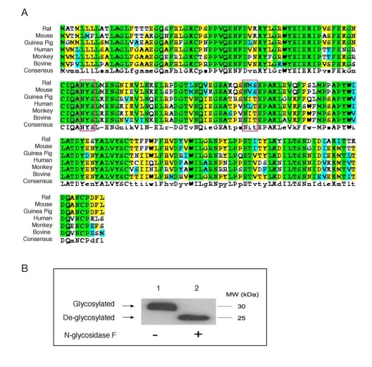
Figure 1.Conservation of N-glycosylation sites in apoD among species. (A). ApoD protein sequences of
different species were aligned using the ClustalW program. Amino acid
residues in box denote two highly conserved N-glycosylation sites in apoD.
The consensus N-glycosylation site is Asn-X-Ser/Thr. (B) Plasma apoD is
N-glycosylated. Aliquots of plasma (20 μg protein) from C57BL/6J mice were
incubated without (-) and with (+) 1,000 U of N-glycosidase F (New England
Biolabs) in a total volume of 30 μl at 37°C for 1 hour to remove N-glycan chains from glycopeptides. The
reaction mixture was resolved on 4-20% SDS-polyacrylamide gels, followed by
immunoblot analysis using anti-apoD. Glycosylated and de-glycosylated forms
of apoD are indicated.