Correction|Volume 15, Issue 24|pp 15703—15704 Correction for: Sonic hedgehog signaling promotes angiogenesis of endothelial progenitor cells to improve pressure ulcers healing by PI3K/AKT/eNOS signaling
Jianhua Wang1Department of Orthopaedics, Jinan Central Hospital, Jinan, Shandong Province, China, Hongyan Zhan2Department of B-Ultrasound, Fourth People’s Hospital of Jinan, Jinan, Shandong Province, China, Mingming Wang3Department of Orthopaedics, Tengzhou Central People’s Hospital, Tengzhou, Shandong Province, China, Hua Song3Department of Orthopaedics, Tengzhou Central People’s Hospital, Tengzhou, Shandong Province, China, Jianhua Sun3Department of Orthopaedics, Tengzhou Central People’s Hospital, Tengzhou, Shandong Province, China, Gang Zhao1Department of Orthopaedics, Jinan Central Hospital, Jinan, Shandong Province, China
- 1Department of Orthopaedics, Jinan Central Hospital, Jinan, Shandong Province, China
- 2Department of B-Ultrasound, Fourth People’s Hospital of Jinan, Jinan, Shandong Province, China
- 3Department of Orthopaedics, Tengzhou Central People’s Hospital, Tengzhou, Shandong Province, China
Received: November 8, 2023Accepted: November 29, 2023Published: December 29, 2023
Copyright: © 2023 Wang et al. This is an open access article distributed under the terms of the Creative Commons Attribution License (CC BY 4.0), which permits unrestricted use, distribution, and reproduction in any medium, provided the original author and source are credited.
This article has been corrected: The authors found that the image of the control endothelial progenitor cells (EPCs) in the tube formation assay shown in Figure 3A was incorrect due to their unintentional reuse of the image of cells treated with the PI3K inhibitor Y294002. They replaced the incorrect image with an image of control cells from the same experiment. This correction has no impact on the experimental outcome or conclusions.
The corrected Figure 3 is shown below.
The authors would also like to update the affiliation and contact information as follows:
Jianhua Wang1,3, Hongyan Zhan2, Mingming Wang3, Hua Song3, Jianhua Sun3, Gang Zhao1
1Department of Orthopaedics, Jinan Central Hospital, Shandong University, Jinan, Shandong Province, China
2Department of B-Ultrasound, Fourth People’s Hospital of Jinan, Jinan, Shandong Province, China
3Department of Orthopaedics, Tengzhou Central People’s Hospital, Tengzhou, Shandong Province, China
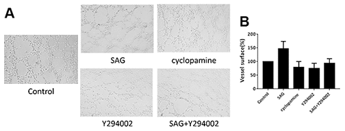
Figure 3. SHH pathway induces angiogenesis properties of EPCs by PI3K/AKT/eNOS signaling. (A, B) The EPCs were treated with SAG (1 μM), cyclopamine (10 μM), Y294002 (5 μM), or co-treated with SAG (1 μM) and Y294002 (5 μM). The angiogenesis properties were analyzed by tube formation assays. N = 3.


