


Introduction
Bone metabolism is a dynamic process that involves the simultaneous processes of bone resorption and formation [1, 2] Thus, osteoblast and osteoclast disequilibrium can undermine bone integrity and normal functionality, promoting a series of pathological lytic bone disorders [3–5]. Thus, recent research has centered on the investigation and identification of novel agents (synthetic or natural) which may inhibit the pathological formation of osteoclasts and bone resorption [6, 7].
Osteoclasts (OCs) are a type of multinucleated giant cell derived from macrophage precursors. OC differentiation is the first stage of the bone resorptive process, which requires stimulation of receptor activator of NF-κB ligand (RANKL) and macrophage colony stimulating factor (M-CSF). Previous studies have shown that RANKL plays an essential role in the formation of multinucleated OCs [8, 9]. Following an interaction between RANKL and receptor activator of NF-κB (RANK), various downstream signaling pathways (e.g., MAPKs and PI3K/Akt) are activated. These specific pathways are able to upregulate the key regulator of the osteoclast process: nuclear factor of activated T cells c1 (NFATc1) [10–13].
In previous studies, Notch signaling has been shown to play an essential role in multiple developmental pathways [14]. The interaction between Notch receptors and Delta-like1/3/4 (Dlk1/3/4) proteins causes the release of tumor necrosis factor α-converting enzyme (TACE), and γ-secretase [15]. Previous studies have confirmed that the down-regulation of γ-secretase has an inhibitory effect on Notch receptor cleavage. Moreover, Notch has been reported to exhibit both an enhancive and prohibitive effect on RANKL-induced bone resorption [16], suggesting that it may represent a promising targetable pathway for the development of anti-resorptive agents [17, 18]. Furthermore, RO4929097 is a γ-secretase inhibitor that suppresses Notch cleavage; however, its effect on the regulation of osteoclast in the regulation of osteoclast formation has not been confirmed.
In this study, we investigated the effect of RO4929097 on the suppression of RANKL-induced osteoclast formation and elucidated the potential molecular mechanisms associated with this process. We confirmed that RO4929097 suppressed osteoclastogenesis and bone resorption by blocking the Notch/Hes1/MAPK (Erk and p38)/Akt pathway. This conclusion was also confirmed by the in vivo results.
Materials and Methods
BMMs and culture system
BMMs were obtained and cultured as described in previous studies [19–21]. Primary BMMs were isolated from the femurs and tibiae of five-week-old male C57/BL6 mice. All processes were performed under the supervision of Shanghai General Hospital Animal Center Committee of Animal Care and Use (2018371A218). All procedures were conducted in accordance with the guidelines of the Ethical Conduct in the Care and Use of Nonhuman Animals in Research. To select primary BMMs, cells were cultured in complete α-MEM (10% Gibco FBS) containing 33.3 ng/mL M-CSF (R&D Systems, Inc.).
Cell cytotoxicity assay
The cytotoxicity of RO4929097 (Selleck, Inc.) was determined using an MTT assay. The BMMs were seeded into 96-well plates (1 × 10⁴ cells per well) in complete medium containing 33.3 ng/mL M-CSF and different concentrations of RO4929097 (0, 0.1, 0.2, 0.4, 0.8, 1.6, 3.2, 6.4, 12.8, and 25.6 μM). Following RO4929097 treatment, the cells were incubated for different timepoints (24 h, 48 h, and 96 h). At the end of the process, CCK-8 solution (Dojindo Molecular Technologies, Inc.) was used according to according to the manufacturer’s guidelines.
Osteoclast formation and TRAP staining assay
The role of RO4929097 on osteoclast formation was assessed. The BMMs were seeded into 96-well plates (1 × 10⁴ cells per well) and stimulated with RANKL (100 ng/mL, R&D Systems, Inc.), M-CSF (33.3 ng/mL), and different concentrations of RO4929097 (0, 100, 200, and 400 nM). The culture medium was replaced every 48 h for six days. After the osteoclasts were observed in the control group, 4% paraformaldehyde was used for OC fixation. TRAP (Sigma-Aldrich; Merck, Inc.) staining solution was added into each of the wells at 37°C for 1 h. TRAP+ cells with at least three nuclei were considered to be osteoclasts. Image J software (National Institutes of Health) was used for cell enumeration.
Bone resorption assay
M-CSF-dependent BMMs were incubated in six-well plates (20 × 105 cells/well). After 24 h, the cells were stimulated with RANKL (100 ng/mL), M-CSF (33.3 ng/mL) until OC formation was observed. The OCs (1 × 10⁴ cells/well ) were then incubated on Corning Osteo Assay Surface plates (Corning, Inc.) and cultured with M-CSF (33.3 ng/mL), RANKL (100 ng/mL), and RO4929097 (0, 100, 200, and 400 nM) for four days. A BioTek Cytation 3 Cell Imaging Reader (BioTek, Winooski, VT) and Image J software were used to photograph and analyze the total resorption pits.
Immunofluorescence analysis of the podosomal actin belt
OCs derived from BMMs were generated and processed. On days 5–7, pancake-like osteoclasts were observed in the RANKL-treated control group, fixed with 0.1% Triton X-100 (Sigma-Aldrich; Merck&Co, Inc.), and permeabilized for 5 min. After blocking with 1% BSA-PBS for 1 h, the cytoskeletal actin structure was stained with rhodamine-conjugated phalloidin. The immunofluorescence images were obtained using a BioTek Cytation 3 Cell Imaging Reader. Image J software was used to analyze the size (diffusion area) and number of the dental actin bands.
Real-time PCR analysis
The total RNA (2 × 10⁶ cells) was extracted from the cells using TRIzol® (Invitrogen; Thermo Fisher Scientific, Inc.) according to the manufacturer's protocol. A NanoDrop 2000 spectrophotometer (NanoDrop Technologies; Thermo Fisher Scientific, Inc.) was used to assess the concentration of the total RNA. The following process was performed as described previously. [22] The following mouse primer sets were used in accordance with published reports [21, 23]: mouse NFATc1: forward, 5′-TGCTCCTCCTCCTGCTGCTC-3′ and reverse, 5′-GCAGAAGGTGGAGGTGCAGC-3′; mouse C-fos: forward, 5′-CCAGTCAAGAGCATCAGCAA-3′ and reverse, 5’-AAGTAGTGCAGCCCGGAGTA-3′; mouse Cath-K: forward, 5′-CTTCCAATACGTGCAGCAGA-3′ and reverse, 5′-TCTTCAGGGCTTTCTCGTTC-3′; mouse Trap: forward, 5′-CAAAGAGATCGCCAGAACCG-3′ and reverse, 5′-GAGACGTTGCCAAGGTGATC-3’; mouse GAPDH: forward, 5′-CACCATGGGA GAAGGCCGGGG-3′ and reverse, 3′-GACGGACACA TTGGGGGTAG-5′.
Western blotting analysis
The total cellular proteins (TCPs) were extracted at different time points. Following treatment with 400 nM RO4929097 for 2 h, RANKL (100 ng/mL) was used to stimulate the cells for different periods of time (short time course). The membrane was blocked in 1% TBST (tri-buffered saline, Tween 20) in 5% skim milk at room temperature for 1 h, and subsequently reacted with primary antibodies NFATc1 (cat no. 5861S; 1:1,000; Cell Signaling Technology, Inc.); Cleaved Notch1 (cat no. 4147S; 1:1,000; Cell Signaling Technology, Inc.); HES1 (cat no.11988; 1:1,000; Cell Signaling Technology, Inc.); GAPDH (cat no.174S; 1:1,000; Cell Signaling Technology, Inc.); p-Akt (cat no.4060S; 1:1,000; Cell Signaling Technology, Inc.); Akt (cat no.4685S; 1:1,000; Cell Signaling Technology, Inc.); p-ERK (cat no.4370S; 1:1,000; Cell Signaling Technology, Inc.); Erk (cat no.9194S; 1:1,000; Cell Signaling Technology, Inc.); p65 (cat no.8242S; 1:1,000; Cell Signaling Technology, Inc.); p-p38 (cat no.4511; 1:1,000; Cell Signaling Technology, Inc.); p38 (cat no.8690; 1:1,000; Cell Signaling Technology, Inc.); p-p65 (cat no.3033; 1:1,000; Cell Signaling Technology, Inc.). After incubating the samples overnight at 4°C, they were incubated with a secondary antibody at room temperature for 1 h using Odyssey V3 .0 image scan (Li-COR. Inc., Lincoln, NE, USA) to observe antibody reactivity.
Mouse model of LPS-Induced calvarial bone resorption
All processes were was carried out in compliance with the ARRIVE guidelines and under the supervision of Shanghai General Hospital Animal Center Committee of Animal Care and Use. All procedures were performed under the guidelines of the Ethical Conduct in the Care and Use of Nonhuman Animals in Research. Male C57BL/6 mice (aged 7 to 8 weeks), were randomly assigned to four groups: Control (PBS), LPS and LPS with low (400 nM) or high (800 nM) concentrations of RO4929097. A 1% pentobarbital sodium (10 mg/kg) was injected intraperitoneally to anaesthetize the mice. The knees of the mice were locally injected with 100 μL LPS or PBS (10 mg/kg), and with or without RO4929097, respectively, for two weeks. During the experiment period, no significant adverse or toxic effects were observed. The mice did not exhibit significant weight loss or lack of spirit, and none of the mice died during the experimental period. Then, euthanasia was performed using pentobarbital sodium (100 mg/kg) [24]. The knee joints were resected and fixed in 4% paraformaldehyde for 48 h. Micro-computed tomography (CT) scanning was performed using high-resolution micro-CT (μCT-100, SCANCO Medical AG, Switzerland). The resolution of the scanning was 10 μm; the X-ray energy was set at 70 kv, 200 μA; and the fixed exposure time was 300 ms. We defined the region of interest (ROI) to encompass the entire subchondral bone in the tibial plateaus.
Histological staining and histomorphometric analysis
The PFA-fixed mice knees were decalcified in 10% EDTA for two weeks and embedded in paraffin. Next, the samples were sliced into specific sections (4 μm thick) and subjected to H&E and TRAP staining. Digital images were obtained using an Axio ScopeA1 light microscope (ZEISS, Inc.). The osteoclasts were enumerated using Image J software.
Statistical analysis
All data were presented as the mean ± standard deviation (SD). A Student's t-test was used to assess the differences between the control and therapeutic groups. The results for the multiple group comparisons were analyzed using a Scheffe's test and one-way analysis of variance (ANOVA) with SPSS 22.0 software (SPSS, Inc.). Values were determined to be significant at *P < 0.05; **P < 0.01; and ***P < 0.001.
Availability of data and materials
The datasets supporting the conclusions of this article are available in an open access repository.
Ethics approval and consent to participate
The study design was carried out in compliance with the ARRIVE guidelines and approved by the Ethical Committee of Shanghai General Hospital.
Results
RANKL-induced osteoclast formation in vitro was down-regulated by RO4929097
First, the cytotoxicity of RO4929097 was evaluated. The results showed that the IC50 of RO4929097 in the BMMs at 24 h, 48 h, and 96 h was 4.44 μM, 3.66 μM, and 2.93 μM respectively (Figure 1A and 1B). Figure 1C presents the chemical structure formula of RO4929097 (provided by Selleck). Compared with the control group, RO4929097 treatment could effectively reduce the number and area of OCs in a dose-dependent manner (Figure 1D, 1E and 1F). Accordingly, RO4929097 was able to inhibit osteoclast formation at a specific concentration under IC50.
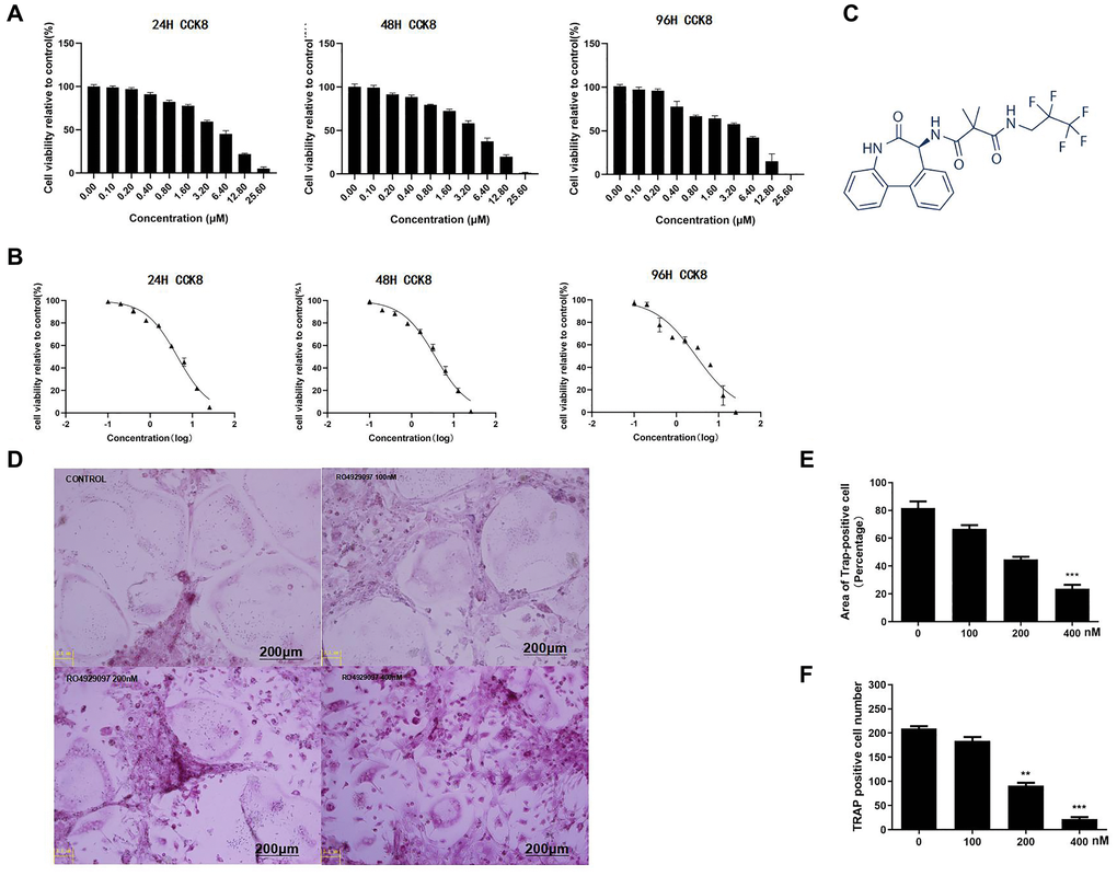
Figure 1. RANKL-Induced osteoclast formation in vitro was down-regulated by RO4929097. (A) RO4929097cytotoxicity. (B) The results showed that the IC50 of RO4929097 at different time points in the BMMs was 4.44 μM, 3.66 μM, and 2.93 μM, respectively. (C) The chemical structural formula of RO4929097 (provided by Selleck). (D) BMMs were simulated with different concentrations of RO4929097 combined with M-CSF (33.3 ng/mL) and RANKL (100 ng/mL) for five days. Cells were fixed in 4% paraformaldehyde and stained for TRAP. (E and F) The area and number of TRAP-positive cells. The data were presented as the mean ± SD (*p < 0.05, **p < 0.01, ***p < 0.001).
Podosome actin belt formation and OC-mediated bone resorption activity was inhibited by RO4929097
To assess the effect of RO4929097 on OC-mediated bone resorption activity, the resorption area on the hydroxyapatite-coated Osteo Assay plates was calculated (Figure 2A). In accordance with the observed osteoclast reduction, the bone resorption area was also reduced in a dose-dependent manner (Figure 2B). The immunofluorescence results showed that podosome actin belt formation was suppressed by RO4929097 in a dose-dependent manner (Figure 2C). In summary, these data show that RO4929097 could effectively inhibit podosome actin belt formation (Figure 2D).

Figure 2. Podosome actin belt formation and OC-mediated bone resorption activity was inhibited by RO4929097. (A) OCL were seeded onto Osteo Assay Stripwell Plates with RANKL (100 ng/mL), M-CSF (33.3 ng/mL), and RO4929097 (0,100, 200, and 400 nM) for four days (B) The resorption area in Osteo Assay Stripwell Plates. (C) BMMs were cultured in 48-well plates with 0, 100, 200, and 400 nM RO4929097 for five days. The cells were fixed and stained for immunofluorescence. (D) Area of OCs with an F-actin belt. The data were presented as the mean ± SD.
RO4929097 suppressed the relative expression of osteoclastogenesis genes
Quantitative PCR (qPCR) was used to evaluate the relative expression of osteoclastogenesis genes at the mRNA level. It was observed that the relative gene expression of NFATc1, C-fos, Cath-K, and TRAP was markedly downregulated by RO4929097 in both a dose-dependent and time-dependent manner. (Figure 3A and 3B).
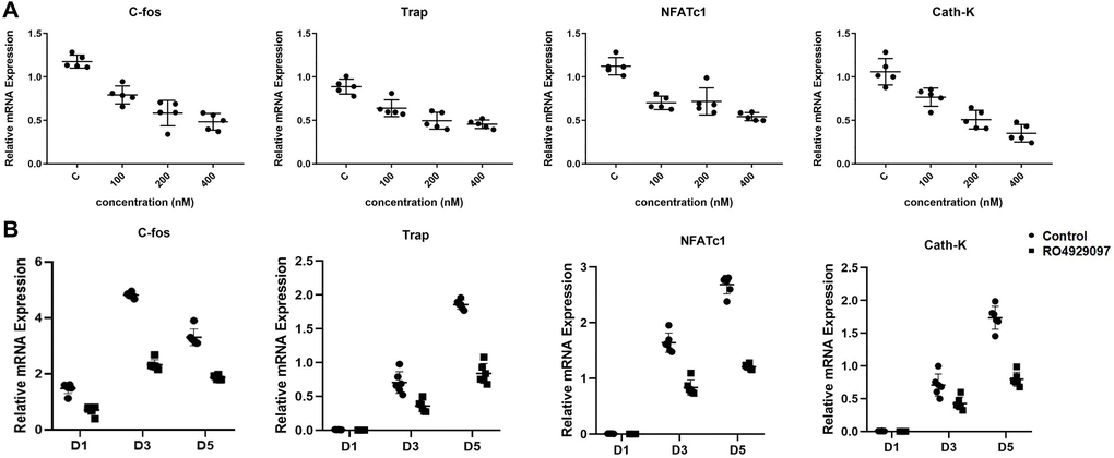
Figure 3. RO4929097 suppressed the relative expression of osteoclastogenesis genes. (A) BMMs were treated with M-CSF (33.3 ng/mL), RANKL (100 ng/mL) in the presence of 0, 100, 200, and 400 nM RO4929097 for five days. The expression of the osteoclast-specific genes, including NFATc1, Cath-K, TRAP, and C-fos were analyzed using quantitative real-time PCR. (B) The BMMs were treated with M-CSF (33.3 ng/mL) and RANKL (100 ng/mL) in the presence 400 nM RO4929097 for 1, 3, and 5 days. Osteoclast-specific gene expression was analyzed using quantitative real-time PCR. RNA expression levels were normalized to the expression of GAPDH. The data were presented as the mean ± SD (*p < 0.05; **p < 0.01; ***p < 0.001).
RO4929097 mediates osteoclastogenesis by inhibiting Notch, MAPK, and Akt signaling in BMMs
To investigate the potential molecular mechanisms of RO4929097 in the inhibition osteoclastogenesis, the RANKL-induced NF-κB, MAPK, JNK and PI3K/Akt pathways were examined using a Western blot analysis. BMM treatment with RO4929097 during RANKL-induced osteoclast formation induced cleaved Notch1, Hes1, and NFATc1 protein expression (Figure 4A and 4B). In addition, short-term stimulation with RO4929097 significantly reduced the phosphorylation of p38, ERK1/2, p65, JNK and Akt (Figure 4C). Accordingly, our results suggest that RO4929097 inhibited osteoclastogenesis by preventing Notch receptor cleavage and Notch signaling, as well as by perturbing the activation of the ERK1/2, p38, JNK and AKT signaling cascades.
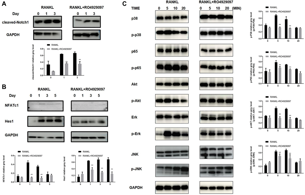
Figure 4. RO4929097 suppresses osteoclastogenesis via Notch inhibition and the regulation of AKT phosphorylation. (A) RO4929097 treatment suppressed the expression of cleaved Notch1. (B) RO4929097 treatment suppressed the protein expression of Hes1 and NFATc1. (C) RO4929097 treatment suppressed the expression of AKT phosphorylation. The data were presented as the mean ± SD (*p < 0.05; **p < 0.01; ***p < 0.001).
RO4929097 administration prevents LPS-induced bone resorption in vivo
The in vitro experiments were used to investigate the effect of RO4929097 on RANKL-induced osteoclastogenesis and bone resorption function by studying the phenotype and associated mechanism. We investigated the function of RO4929097 in mice with LPS-induced bone resorption. According to the three-dimensional micro-CT reconstruction shown in Figure 5A, compared with the control group, the RO4929097-treated groups displayed fewer and smaller resorption pits. A significant reduction in BV/TV, Tb.n, and BMD was also observed in the LPS group (Figure 5B).
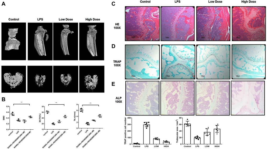
Figure 5. RO4929097 attenuated lipopolysaccharide-induced bone resorption in mouse knees in vivo. (A) Images of a three dimension reconstruction based on micro-CT scanning. (B) Results of BMD, bone BV/TV and Tb.n. (C, D and E) HE TRAP and ALP results. Amount of TRAP-positive OCs. The data are presented as the mean ± SD (*p < 0.05; **p < 0.01; ***p < 0.001).
The histological analysis also revealed an inhibitory effect of RO4929097 on LPS-induced bone resorption in vivo. In accordance with previous results, extensive osteolysis was observed in the HE-stained images of the control group, whereas a lower osteolytic level was observed in the RO4929097-treated groups (Figure 5C). The TRAP staining results confirmed a decreased number of OCs following RO4929097 treatment (Figure 5D). No difference was observed in the ALP staining under RO4929097 treatment (Figure 5E). Accordingly, the in vivo results confirmed the potential therapeutic application of RO4929097 in inflammation-relative osteolytic disease.
Discussion
Pathological bone homeostasis promotes the over-formation and activation of OCs, leading to substantial bone disruption, which is a feature of several osteolytic diseases [3–5]. Medical therapies that suppress osteolysis are considered to be a potential treatment strategy for these diseases [25, 26]. However, several of these treatments are associated with various side-effects [27, 28]. Accordingly, the development of medications that can inhibit osteoclastogenesis without side-effects are urgently required.
Our research reveals potential signaling pathways which may regulate RANKL-induced osteoclast formation. Notch signaling is important in many cellular functions, including morphogenesis and stem cell niche maintenance [29–31]. However, the function of Notch signaling on osteoclastogenesis remains unclear, with evidence supporting both a stimulatory and inhibitory role. According to previous research, Notch has been shown to act in conjunction with MAPK, NF-κB, and PI3K/Akt to potentially regulate NFATc1 expression during osteoclast differentiation [17, 32–35]. In addition, Notch1 suppression in BMMs has been confirmed to promote osteoclastogenesis in vitro [36], whereas Notch2 suppression attenuates osteoclastogenesis [16, 37].
Previous research has confirmed a synergistic function between Notch and NF-κB signaling [38]. Our findings indicate that NFATc1 expression was significantly decreased in response to RO4929097 treatment. As a potent γ-secretase inhibitor, RO4929097 does not alter the level of Notch expression but rather blocked its cleavage. Accordingly, the expression of cleaved Notch1 (Notch intracellular domain-1; NICD1) and hairy enhancer of split 1 (HES1), a targeted gene regulated by cleaved Notch1, were evaluated were evaluated in BMMS stimulated for longer time points. In accordance with previous studies [39], we also observed a reduction in the level of ERK, p38, Akt, and p65 phosphorylation in response to RO4929097 treatment. This indicates that there is significant crosstalk between Notch, ERK, and Akt signaling. Our data do suggest that RO4929097 can suppress bone loss and osteoclast formation via Notch-mediated NFATc1 expression.
In this study, a widely-accepted LPS-induced osteolytic experimental model was established to assess the effect of RO4929097. During the experimental design for this study, Ovx, RANKL-induced bone resorption, and LPS- induced bone resorption models were considered. The Ovx model was considered to be inappropriate for this research because Ovx regulates both osteoclast and osteoblast formation. The reason RANKL is used for in vitro studies is because RANKL is mainly produced by osteoblasts and is necessary for the formation of osteoclasts; however, osteoblasts are present in vivo. LPS-induced bone resorption, as a general approach, has been widely used for osteoclast-related in vivo studies. [40–44] Thus, LPS-induced bone resorption was selected as the in vivo model in this study. Both the micro-CT analysis and histological staining of TRAP confirmed downregulated osteopenia in the RO4929097-treated groups. However, the present study was associated with some limitations. First, our study did not address the possibility that RO4929097 regulates osteoclasts via other signaling pathways. Second, the influence of RO4929097 on osteoblast and bone formation should be further assessed.
In conclusion, our data demonstrate that RO4929097 can effectively attenuate RANKL-induced osteoclastogenesis in vitro via downregulating Notch/MAPK (ERK and p38)/Akt signaling. In accordance with the in vitro observations, prevention of LPS-induced osteolysis in vivo also confirmed that RO4929097 may represent a potential treatment strategy for osteolytic diseases in relation to excessive osteoclast-mediated bone resorption.
Author Contributions
Donghua Hang was responsible for data analysis and interpretation. Lei Wang and Yuan Zhou recorded and analyzed the in vivo data. Yi Zhao built the 3-D model. Congyunn Zhao performed the animal experiment and collected the samples. Tao Huang is the first authors who performed all the in vitro experiment and responsible for the conception and critical revision of the article for important intellectual content. All of the authors read and approved the manuscript.
Acknowledgments
This study was financially supported by the National Natural Science Foundation of China (grant nos. 81201439).
Conflicts of Interest
The authors declare they have no conflicts of interest.
Funding
This study was financially supported by the National Natural Science Foundation of China (grant nos. 81201439).
References
- 1. Langdahl B, Ferrari S, Dempster DW. Bone modeling and remodeling: potential as therapeutic targets for the treatment of osteoporosis. Ther Adv Musculoskelet Dis. 2016; 8:225–35. https://doi.org/10.1177/1759720X16670154 [PubMed]
- 2. Rachner TD, Khosla S, Hofbauer LC. Osteoporosis: now and the future. Lancet. 2011; 377:1276–87. https://doi.org/10.1016/S0140-6736(10)62349-5 [PubMed]
- 3. Bertuglia A, Lacourt M, Girard C, Beauchamp G, Richard H, Laverty S. Osteoclasts are recruited to the subchondral bone in naturally occurring post-traumatic equine carpal osteoarthritis and may contribute to cartilage degradation. Osteoarthritis Cartilage. 2016; 24:555–66. https://doi.org/10.1016/j.joca.2015.10.008 [PubMed]
- 4. Mukherjee K, Chattopadhyay N. Pharmacological inhibition of cathepsin K: A promising novel approach for postmenopausal osteoporosis therapy. Biochem Pharmacol. 2016; 117:10–19. https://doi.org/10.1016/j.bcp.2016.04.010 [PubMed]
- 5. Kapasa ER, Giannoudis PV, Jia X, Hatton PV, Yang XB. The Effect of RANKL/OPG Balance on Reducing Implant Complications. J Funct Biomater. 2017; 8:42. https://doi.org/10.3390/jfb8040042 [PubMed]
- 6. Alami S, Hervouet L, Poiraudeau S, Briot K, Roux C. Barriers to Effective Postmenopausal Osteoporosis Treatment: A Qualitative Study of Patients' and Practitioners' Views. PLoS One. 2016; 11:e0158365. https://doi.org/10.1371/journal.pone.0158365 [PubMed]
- 7. Tella SH, Gallagher JC. Prevention and treatment of postmenopausal osteoporosis. J Steroid Biochem Mol Biol. 2014; 142:155–70. https://doi.org/10.1016/j.jsbmb.2013.09.008 [PubMed]
- 8. Biskobing DM, Fan X, Rubin J. Characterization of MCSF-induced proliferation and subsequent osteoclast formation in murine marrow culture. J Bone Miner Res. 1995; 10:1025–32. https://doi.org/10.1002/jbmr.5650100706 [PubMed]
- 9. Udagawa N, Takahashi N, Jimi E, Matsuzaki K, Tsurukai T, Itoh K, Nakagawa N, Yasuda H, Goto M, Tsuda E, Higashio K, Gillespie MT, Martin TJ, Suda T. Osteoblasts/stromal cells stimulate osteoclast activation through expression of osteoclast differentiation factor/RANKL but not macrophage colony-stimulating factor: receptor activator of NF-kappa B ligand. Bone. 1999; 25:517–23. https://doi.org/10.1016/S8756-3282(99)00210-0 [PubMed]
- 10. Huang H, Ryu J, Ha J, Chang EJ, Kim HJ, Kim HM, Kitamura T, Lee ZH, Kim HH. Osteoclast differentiation requires TAK1 and MKK6 for NFATc1 induction and NF-kappaB transactivation by RANKL. Cell Death Differ. 2006; 13:1879–91. https://doi.org/10.1038/sj.cdd.4401882 [PubMed]
- 11. Novack DV. Role of NF-κB in the skeleton. Cell Res. 2011; 21:169–82. https://doi.org/10.1038/cr.2010.159 [PubMed]
- 12. Strickson S, Emmerich CH, Goh ET, Zhang J, Kelsall IR, Macartney T, Hastie CJ, Knebel A, Peggie M, Marchesi F, Arthur JS, Cohen P. Roles of the TRAF6 and Pellino E3 ligases in MyD88 and RANKL signaling. Proc Natl Acad Sci U S A. 2017; 114:E3481–89. https://doi.org/10.1073/pnas.1702367114 [PubMed]
- 13. Lee K, Chung YH, Ahn H, Kim H, Rho J, Jeong D. Selective Regulation of MAPK Signaling Mediates RANKL-dependent Osteoclast Differentiation. Int J Biol Sci. 2016; 12:235–45. https://doi.org/10.7150/ijbs.13814 [PubMed]
- 14. Lai EC. Notch signaling: control of cell communication and cell fate. Development. 2004; 131:965–73. https://doi.org/10.1242/dev.01074 [PubMed]
- 15. Chen S, Lee BH, Bae Y. Notch signaling in skeletal stem cells. Calcif Tissue Int. 2014; 94:68–77. https://doi.org/10.1007/s00223-013-9773-z [PubMed]
- 16. Sekine C, Koyanagi A, Koyama N, Hozumi K, Chiba S, Yagita H. Differential regulation of osteoclastogenesis by Notch2/Delta-like 1 and Notch1/Jagged1 axes. Arthritis Res Ther. 2012; 14:R45. https://doi.org/10.1186/ar3758 [PubMed]
- 17. Ashley JW, Ahn J, Hankenson KD. Notch signaling promotes osteoclast maturation and resorptive activity. J Cell Biochem. 2015; 116:2598–609. https://doi.org/10.1002/jcb.25205 [PubMed]
- 18. Duan L, de Vos P, Fan M, Ren Y. Notch is activated in RANKL-induced osteoclast differentiation and resorption. Front Biosci. 2008; 13:7064–71. [PubMed]
- 19. Wu C, Wang W, Tian B, Liu X, Qu X, Zhai Z, Li H, Liu F, Fan Q, Tang T, Qin A, Zhu Z. Myricetin prevents titanium particle-induced osteolysis in vivo and inhibits RANKL-induced osteoclastogenesis in vitro. Biochem Pharmacol. 2015; 93:59–71. https://doi.org/10.1016/j.bcp.2014.10.019 [PubMed]
- 20. Zhai Z, Qu X, Li H, Yang K, Wan P, Tan L, Ouyang Z, Liu X, Tian B, Xiao F, Wang W, Jiang C, Tang T, et al. The effect of metallic magnesium degradation products on osteoclast-induced osteolysis and attenuation of NF-κB and NFATc1 signaling. Biomaterials. 2014; 35:6299–310. https://doi.org/10.1016/j.biomaterials.2014.04.044 [PubMed]
- 21. Zhou Z, Chen X, Chen X, Qin A, Mao Y, Pang Y, Yu S, Zhang S. PP121 suppresses RANKL-Induced osteoclast formation in vitro and LPS-Induced bone resorption in vivo. Exp Cell Res. 2020; 388:111857. https://doi.org/10.1016/j.yexcr.2020.111857 [PubMed]
- 22. Chen X, Chen X, Zhou Z, Qin A, Wang Y, Fan B, Xu W, Zhang S. LY411575, a potent γ-secretase inhibitor, suppresses osteoclastogenesis in vitro and LPS-induced calvarial osteolysis in vivo. J Cell Physiol. 2019; 234:20944–56. https://doi.org/10.1002/jcp.28699 [PubMed]
- 23. Chen X, Chen X, Zhou Z, Mao Y, Wang Y, Ma Z, Xu W, Qin A, Zhang S. Nirogacestat suppresses RANKL-Induced osteoclast formation in vitro and attenuates LPS-Induced bone resorption in vivo. Exp Cell Res. 2019; 382:111470. https://doi.org/10.1016/j.yexcr.2019.06.015 [PubMed]
- 24. Laferriere CA, Leung VS, Pang DS. Evaluating Intrahepatic and Intraperitoneal Sodium Pentobarbital or Ethanol for Mouse Euthanasia. J Am Assoc Lab Anim Sci. 2020; 59:264–68. https://doi.org/10.30802/AALAS-JAALAS-19-000097 [PubMed]
- 25. Hiligsmann M, Evers SM, Ben Sedrine W, Kanis JA, Ramaekers B, Reginster JY, Silverman S, Wyers CE, Boonen A. A systematic review of cost-effectiveness analyses of drugs for postmenopausal osteoporosis. Pharmacoeconomics. 2015; 33:205–24. https://doi.org/10.1007/s40273-014-0231-1 [PubMed]
- 26. Hadji P, Papaioannou N, Gielen E, Feudjo Tepie M, Zhang E, Frieling I, Geusens P, Makras P, Resch H, Möller G, Kalouche-Khalil L, Fahrleitner-Pammer A. Persistence, adherence, and medication-taking behavior in women with postmenopausal osteoporosis receiving denosumab in routine practice in Germany, Austria, Greece, and Belgium: 12-month results from a European non-interventional study. Osteoporos Int. 2015; 26:2479–89. https://doi.org/10.1007/s00198-015-3164-4 [PubMed]
- 27. McHorney CA, Schousboe JT, Cline RR, Weiss TW. The impact of osteoporosis medication beliefs and side-effect experiences on non-adherence to oral bisphosphonates. Curr Med Res Opin. 2007; 23:3137–52. https://doi.org/10.1185/030079907X242890 [PubMed]
- 28. Hough FS, Brown SL, Cassim B, Davey MR, de Lange W, de Villiers TJ, Ellis GC, Lipschitz S, Lukhele M, Pettifor JM, and National Osteoporosis Foundation of South Africa. The safety of osteoporosis medication. S Afr Med J. 2014; 104:279–82. https://doi.org/10.7196/SAMJ.7505 [PubMed]
- 29. Artavanis-Tsakonas S, Rand MD, Lake RJ. Notch signaling: cell fate control and signal integration in development. Science. 1999; 284:770–76. https://doi.org/10.1126/science.284.5415.770 [PubMed]
- 30. Bray SJ. Notch signalling: a simple pathway becomes complex. Nat Rev Mol Cell Biol. 2006; 7:678–89. https://doi.org/10.1038/nrm2009 [PubMed]
- 31. Souilhol C, Cormier S, Tanigaki K, Babinet C, Cohen-Tannoudji M. RBP-Jkappa-dependent notch signaling is dispensable for mouse early embryonic development. Mol Cell Biol. 2006; 26:4769–74. https://doi.org/10.1128/MCB.00319-06 [PubMed]
- 32. Hales EC, Taub JW, Matherly LH. New insights into Notch1 regulation of the PI3K-AKT-mTOR1 signaling axis: targeted therapy of γ-secretase inhibitor resistant T-cell acute lymphoblastic leukemia. Cell Signal. 2014; 26:149–61. https://doi.org/10.1016/j.cellsig.2013.09.021 [PubMed]
- 33. Haruki N, Kawaguchi KS, Eichenberger S, Massion PP, Olson S, Gonzalez A, Carbone DP, Dang TP. Dominant-negative Notch3 receptor inhibits mitogen-activated protein kinase pathway and the growth of human lung cancers. Cancer Res. 2005; 65:3555–61. https://doi.org/10.1158/0008-5472.CAN-04-3132 [PubMed]
- 34. Nair P, Somasundaram K, Krishna S. Activated Notch1 inhibits p53-induced apoptosis and sustains transformation by human papillomavirus type 16 E6 and E7 oncogenes through a PI3K-PKB/Akt-dependent pathway. J Virol. 2003; 77:7106–12. https://doi.org/10.1128/JVI.77.12.7106-7112.2003 [PubMed]
- 35. Vacca A, Felli MP, Palermo R, Di Mario G, Calce A, Di Giovine M, Frati L, Gulino A, Screpanti I. Notch3 and pre-TCR interaction unveils distinct NF-kappaB pathways in T-cell development and leukemia. EMBO J. 2006; 25:1000–08. https://doi.org/10.1038/sj.emboj.7600996 [PubMed]
- 36. Bai S, Kopan R, Zou W, Hilton MJ, Ong CT, Long F, Ross FP, Teitelbaum SL. NOTCH1 regulates osteoclastogenesis directly in osteoclast precursors and indirectly via osteoblast lineage cells. J Biol Chem. 2008; 283:6509–18. https://doi.org/10.1074/jbc.M707000200 [PubMed]
- 37. Fukushima H, Nakao A, Okamoto F, Shin M, Kajiya H, Sakano S, Bigas A, Jimi E, Okabe K. The association of Notch2 and NF-kappaB accelerates RANKL-induced osteoclastogenesis. Mol Cell Biol. 2008; 28:6402–12. https://doi.org/10.1128/MCB.00299-08 [PubMed]
- 38. Espinosa L, Inglés-Esteve J, Robert-Moreno A, Bigas A. IkappaBalpha and p65 regulate the cytoplasmic shuttling of nuclear corepressors: cross-talk between Notch and NFkappaB pathways. Mol Biol Cell. 2003; 14:491–502. https://doi.org/10.1091/mbc.e02-07-0404 [PubMed]
- 39. Wang Z, Li Y, Sarkar FH. Notch signaling proteins: legitimate targets for cancer therapy. Curr Protein Pept Sci. 2010; 11:398–408. https://doi.org/10.2174/138920310791824039 [PubMed]
- 40. Matsumoto Y, Larose J, Kent OA, Lim M, Changoor A, Zhang L, Storozhuk Y, Mao X, Grynpas MD, Cong F, Rottapel R. RANKL coordinates multiple osteoclastogenic pathways by regulating expression of ubiquitin ligase RNF146. J Clin Invest. 2017; 127:1303–15. https://doi.org/10.1172/JCI90527 [PubMed]
- 41. Grevers LC, de Vries TJ, Everts V, Verbeek JS, van den Berg WB, van Lent PL. Immune complex-induced inhibition of osteoclastogenesis is mediated via activating but not inhibitory Fcγ receptors on myeloid precursor cells. Ann Rheum Dis. 2013; 72:278–85. https://doi.org/10.1136/annrheumdis-2012-201568 [PubMed]
- 42. Chen YH, Xu DX, Wang JP, Wang H, Wei LZ, Sun MF, Wei W. Melatonin protects against lipopolysaccharide-induced intra-uterine fetal death and growth retardation in mice. J Pineal Res. 2006; 40:40–47. https://doi.org/10.1111/j.1600-079X.2005.00274.x [PubMed]
- 43. Miyaura C, Inada M, Matsumoto C, Ohshiba T, Uozumi N, Shimizu T, Ito A. An essential role of cytosolic phospholipase A2alpha in prostaglandin E2-mediated bone resorption associated with inflammation. J Exp Med. 2003; 197:1303–10. https://doi.org/10.1084/jem.20030015 [PubMed]
- 44. Abu-Amer Y, Ross FP, Edwards J, Teitelbaum SL. Lipopolysaccharide-stimulated osteoclastogenesis is mediated by tumor necrosis factor via its P55 receptor. J Clin Invest. 1997; 100:1557–65. https://doi.org/10.1172/JCI119679 [PubMed]