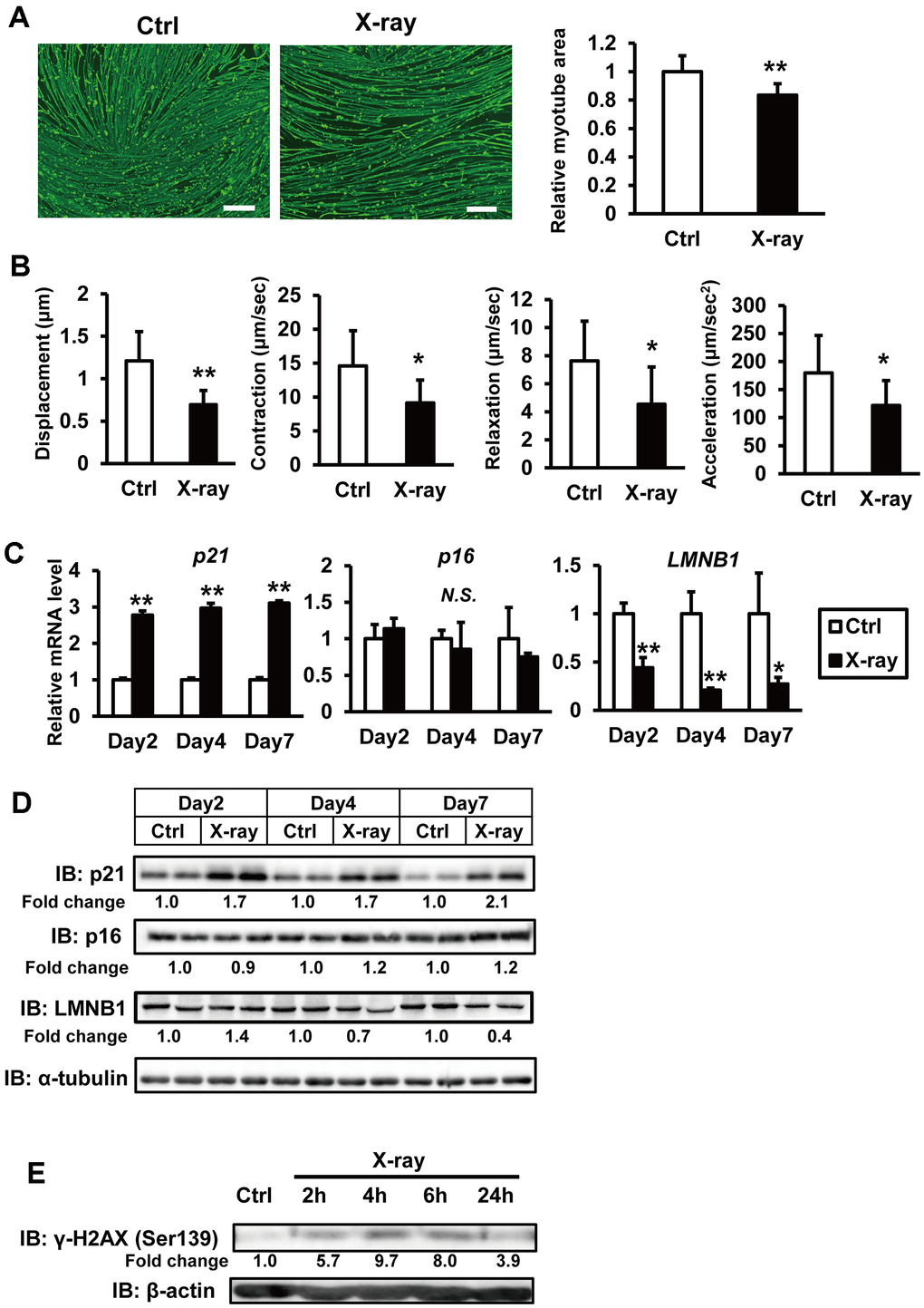
Figure 1.X-ray irradiation induces senescence in hiPSC-derived myocytes. (A) hiPSCs 409B2tet-MyoD were differentiated into myocytes and irradiated with X-rays (3 Gy). One week post-irradiation, cells were fixed, permeabilized, and stained for MHC and nuclei. Scale bar: 100 μm. Relative MHC-positive areas were quantified as described in the methods section. The relative myotube area in nonirradiated controls (Ctrl) was set to 1. (B) hiPSC-derived myocytes were stimulated with electrical pulses (23 V, 4 ms, 1 Hz). Myotube movement was measured using motion analysis software. (C) hiPSC-derived myocytes were irradiated with X-rays (3 Gy), and total RNA was isolated one week later. mRNA levels were measured by real-time PCR and normalized to 18S rRNA. Relative mRNA expression in nonirradiated controls (Ctrl) was set to 1. (D, E) Whole-cell lysates from hiPSC-derived myocytes were collected at the indicated time points after X-ray irradiation and subjected to immunoblotting to assess the expression of the indicated proteins. The numbers under the lanes indicate fold changes in protein level relative to the control and were standardized against α-tubulin or β-actin. Data are presented as mean ± SD (n = 3 or 9). Statistical significance was determined using Student’s t-test. *p < 0.05, **p < 0.01.