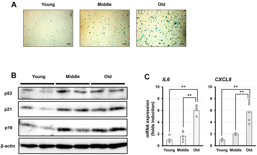
Figure 1.Characterization of senescent cells in vitro. (A) Representative images of senescence-associated β-galactosidase (SA-β-gal) staining in fibroblasts at different senescent stages (young, middle, and old). Scale bar = 200 μm. (B) Representative western blot analysis of p53, p21, and p16, with β-actin as a loading control. (C) PCR analysis of interleukin-6 (IL-6) and interleukin-8 (IL-8) expression. Data are presented as the mean ± standard error (SE) from seven independent experiments (n = 7). Statistical analysis was performed using the Tukey–Kramer test; **p < 0.01.