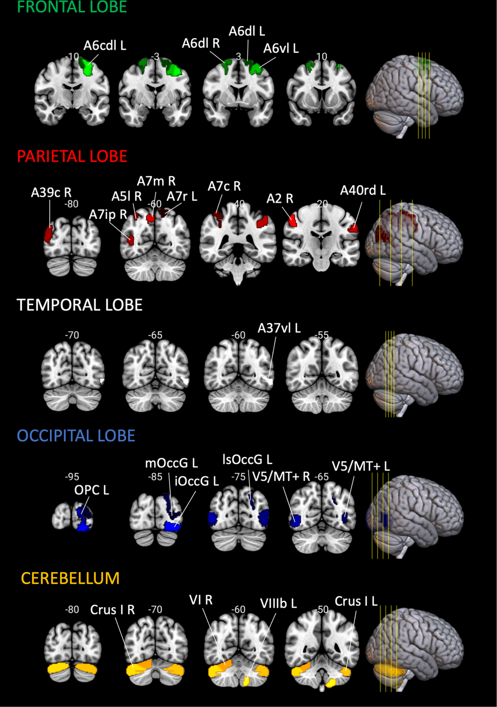
Figure 9.Location of the brain regions analyzed. Brain regions analyzed, visualized in the coronal plane, in the MNI152 Template. Cortical regions were labeled using the Brainnetome Atlas [38], and cerebellar regions with Cerebellum SUIT Atlas [39]. Brain slices and volumes depicted in this figure were created with MRIcroGL (Version 1.2.20220720b; https://www.nitrc.org/projects/mricrogl) [37].