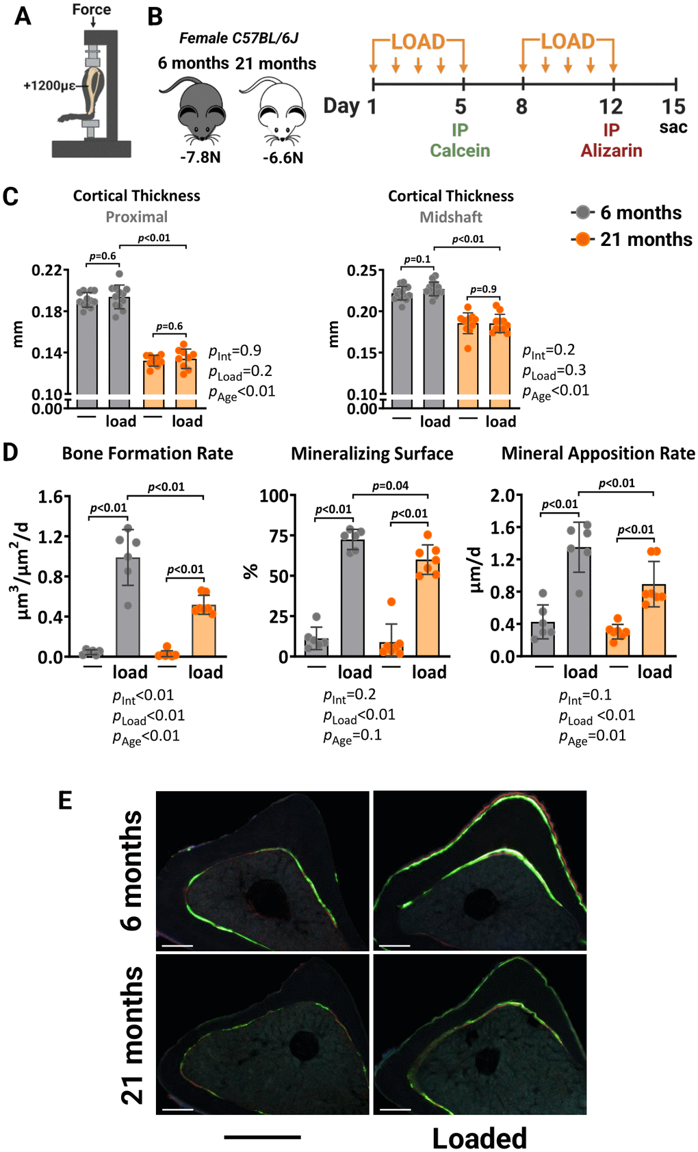
Figure 1.Aging reduces the periosteal osteogenic response to mechanical stimulation in female mice. (A) Schematic illustration of tibia axial compressive loading applied on mouse tibia with +1200με peak strain at the midshaft. (B) Experimental timeline describing the loading and fluorescent dye administration plan. (C) Micro-CT measurements at 5 mm proximal to the tibiofibular junction and at the midshaft of the right (non-loaded) and left (loaded) tibia from young (n=12) and old (n=9) female C57BL/6J mice. (D) Histomorphometric measurements at the periosteal tibial surface 5 mm proximal from the tibiofibular junction of young (n=6) and old (n=7) female mice, and (E) representative photomicrographic cross-section images. Scale bar, 200 μm. Bars represent mean ± SD. Data analyzed using two-way mixed ANOVA; p-values adjusted by Holm-Sidak’s test. Int: Interaction.