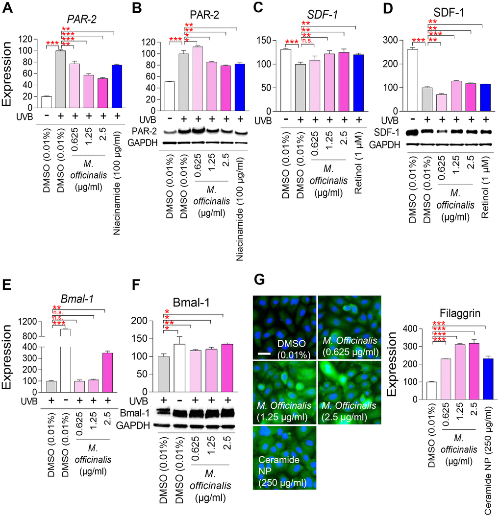
Figure 5.M. officinalis extract reverses skin aging by suppressing skin pigmentation, increasing skin turnover, and maintaining skin moisture. (A, B) Measurement of melanosome transport. To stimulate melanosome production, HaCaT cells were exposed to 30 mJ/cm2 ultraviolet B (UVB). Then, HaCaT cells were treated with DMSO (0.01%) or M. officinalis extract (0.625, 1.25, and 2.5 μg/ml) for 8 h. As a positive control, 100 μg/ml niacinamide was used. PAR–2 expression was analyzed by qPCR (A) and Western blot (B). *P < 0.05, **P < 0.01, ***P < 0.001, Student’s t–test. Mean ± S.D., N = 3. (C, D) Measurement of skin pigmentation. To induce skin pigmentation, HaCaT cells were exposed to 15 J/cm2 UVB. Then, HaCaT cells were treated with DMSO (0.01%) or M. officinalis extract (0.625, 1.25, and 2.5 μg/ml) for 24 h. As a positive control, 1 μM retinol (R7632; Sigma) was used. SDF–1 expression was analyzed by qPCR (C) and Western blot (D). n.s. (not significant), **P < 0.01, ***P < 0.001, Student’s t–test. Mean ± S.D., N = 3. (E, F) Measurement of skin turnover. To disrupt skin turnover, HaCaT cells were exposed to 30 mJ/cm2 UVB. Then, HaCaT cells were treated with DMSO (0.01%) or M. officinalis extract (0.625, 1.25, and 2.5 μg/ml) for 24 h. Bmal–1expression was analyzed by qPCR (E) and Western blot (F). n.s. (not significant), *P < 0.05, **P < 0.01, ***P < 0.001, Student’s t–test. Mean ± S.D., N = 3. (G) Measurement of skin moisture retention. Normal human epidermal keratinocytes, HEKn cells, were treated with DMSO (0.01%) or M. officinalis extract (0.625, 1.25, and 2.5 μg/ml) for 72 h. As a positive control, 250 μg/ml ceramide NP was used. Expression of filaggrin protein was examined using immunocytochemistry (blue: dapi, green: filaggrin). Image J analysis was performed to quantify the fluorescence intensity of filaggrin protein. ***P < 0.001, Student’s t–test. Mean ± S.D., N = 3. The scale bar is 100 μm.