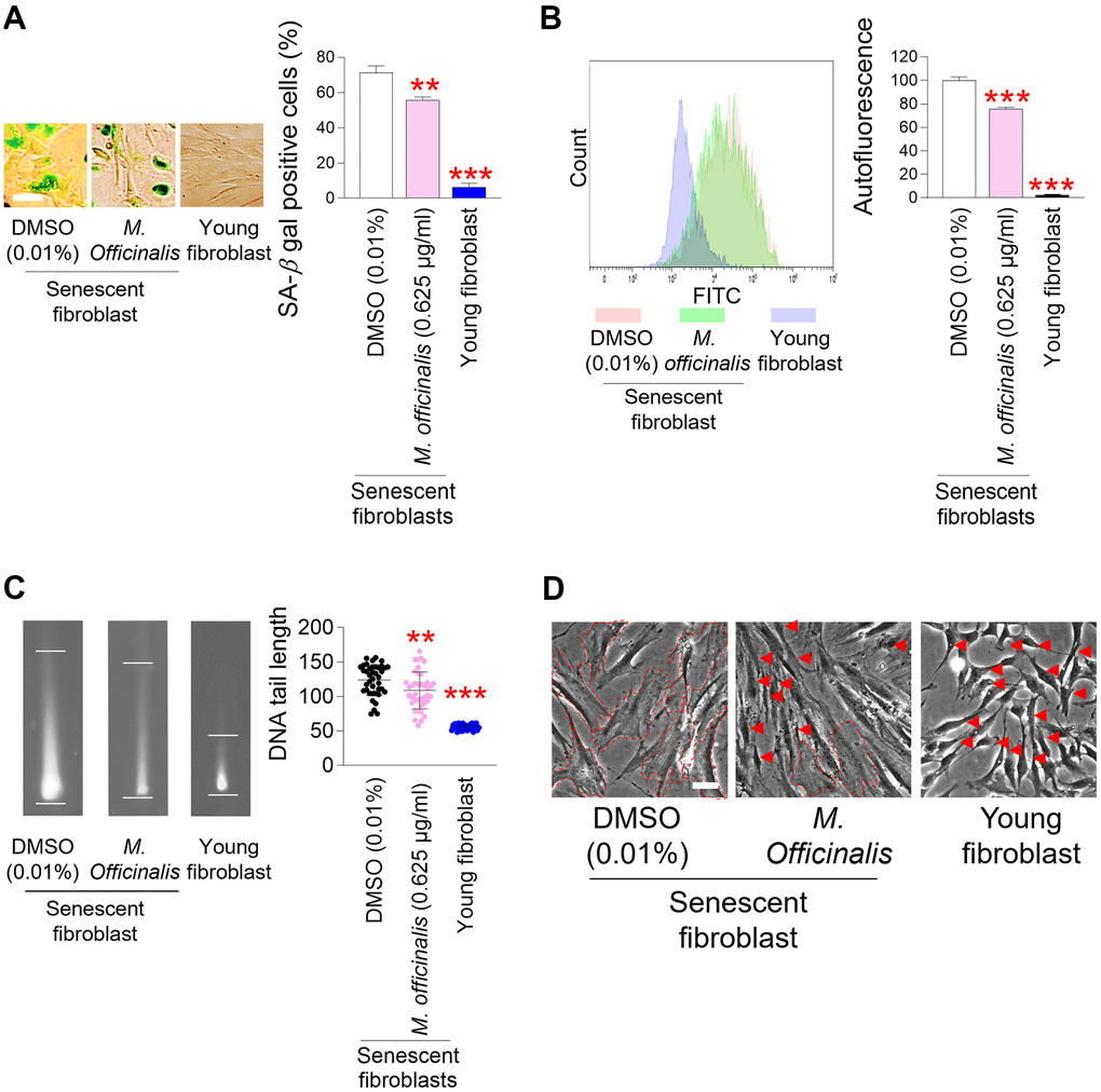
Figure 3.Senescence–associated phenotypes are ameliorated by M. officinalis extract. (A) Measurement of senescence–associated beta–galactosidase (SA–β–gal) positive cells (%). Senescent fibroblasts were treated with DMSO (0.01%) or M. officinalis extract (0.625 μg/ml) for 12 days. Young fibroblasts were used as a positive control. **P < 0.01, ***P < 0.001, Student’s t–test. Means ± S.D., N = 3. Scale bar 20 μm. (B) After 12 days of treatment with DMSO (0.01%) or M. officinalis extract (0.625 μg/ml), autofluorescence was assessed in senescent fibroblasts. Young fibroblasts were used as a positive control. **P < 0.01, ***P < 0.001, Student’s t–test. Means ± S.D., N = 3. (C) After 12 days of treatment with DMSO (0.01%) or M. officinalis extract (0.625 μg/ml), DNA tail length was assessed in senescent fibroblasts by image J. Each dot represents the length of a DNA tail. Young fibroblasts were used as a positive control. **P < 0.01, ***P < 0.001, Student's t–test. Mean ± S.D., N = 40. (D) Morphologies of senescence fibroblasts after 12 days of treatment with DMSO (0.01%) or M. officinalis extract (0.625 μg/ml). Senescent fibroblasts treated with M. officinalis extract exhibited thin and spindly morphology (red arrows), whereas those treated with DMSO exhibited a broad and flat morphology (dotted lines). The scale bar is 20 μm. Young fibroblasts were used as a positive control.