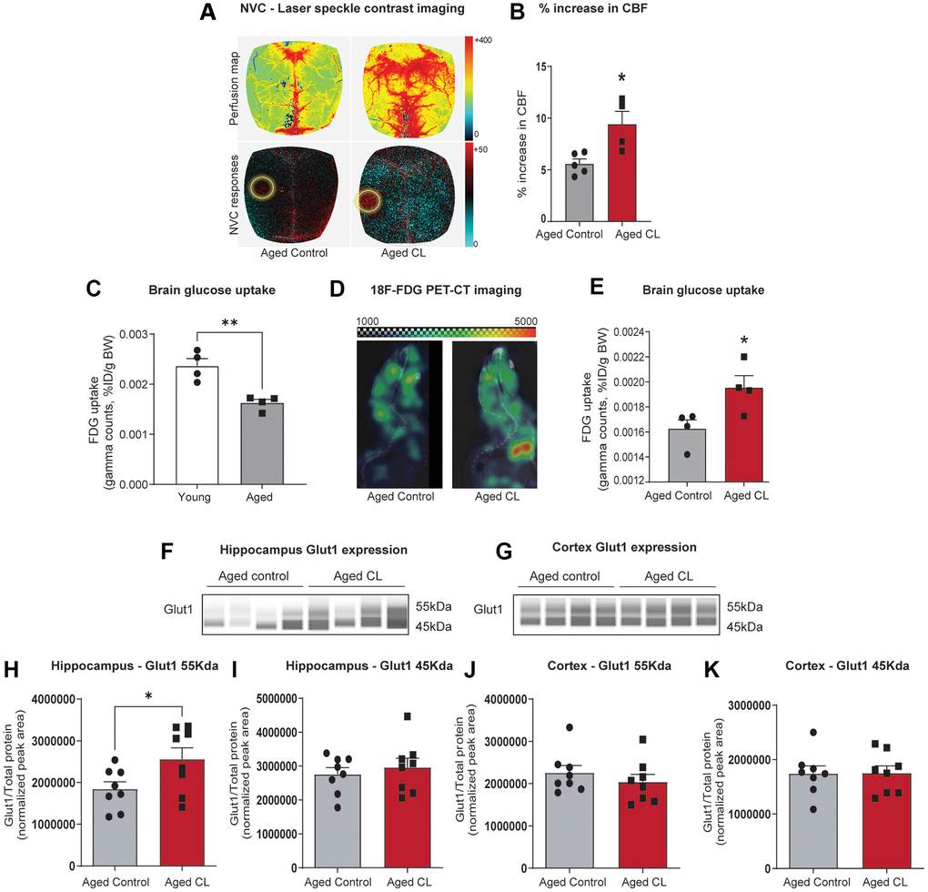
Figure 1.Effects of chronic β3-AR treatment on neurovascular coupling, brain uptake, and GLUT1 expression in aged mice. (A) Representative pseudocolor laser speckle flowmetry maps of baseline cerebral blood flow (CBF) (upper row; shown for orientation purposes) and CBF changes in the somatosensory cortex relative to baseline during contralateral whisker stimulation (bottom row, left circle, 30 s, 5 Hz) in aged mice treated with saline (aged controls) or CL 316,243 (aged CL). The color bar represents CBF as a percent change from the baseline. (B) Summary data as a % increase in CBF (n = 4–5 in each group, males). (C) 18F-FDG uptake in the young and aged brain represented as SUV (%ID/g body weight) (n = 4 in each group, males). (D) Representative 18F-FDG-PET images of aged control and CL-treated mice. Warmer colors represent higher activity in PET images. (E) Quantification of FDG uptake in the brain represented as SUV (%ID/g body weight) (n = 4 in each group, males). (F, G) Representative images of GLUT1 chemiluminescent signals for hippocampus and cortex lysates in capillaries created by the compass SW software for Jess analysis. (H–K) Peak areas for 55 and 45kDa GLUT1 isoforms normalized for total protein in the samples. Data are mean ± S.E.M. (n = 8–9 in each group, males). *P < 0.05 vs. aged controls.