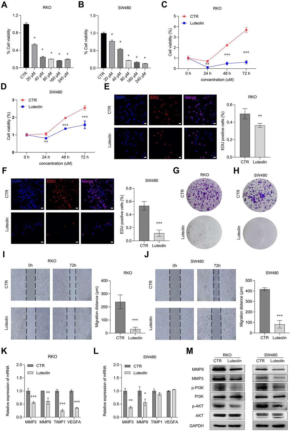
Figure 7.Luteolin represses the malignant phenotypes of CRC cells. (A, B) After treatment with different concentrations of luteolin (0, 20, 40, 80, 160, 200 μM) for 48 h, the cell viabilities of RKO and SW480 cells were determined by using CCK-8 assay. (C, D) After treatment with 40 μM luteolin or DMSO (control) for 24 h, 48 h and 72 h, the cell proliferation capacities of RKO and SW480 cells were determined using CCK-8. (E, F) After treatment with 40 μM luteolin or DMSO (control) for 24 h, the cell proliferation capacities of RKO and SW480 cells were determined using EdU assay. Bar = 20 μm. (G, H) After treatment with 40 μM luteolin or DMSO (control) for 2 weeks, the cell colonies were stained with crystal violet to display the effect of luteolin on proliferation of RKO and SW480 cells. (I, J) After treatment with 40 μM luteolin or DMSO (control) for 72 h, the migration distance of RKO and SW480 cells were assayed by wounding healing experiments. (K, L) After treatment with 40 μM luteolin or DMSO (control) for 24h, the expression level of MMP3, MMP9, TIMP1 and VEGFA in RKO and SW480 cells was detected by qRT-PCR assay. (M) After treatment with 40 μM luteolin or DMSO (control) for 24h, the expression levels of MMP9, MMP3, PI3K, p-PI3K, AKT and p-AKT in RKO and SW480 cells was analyzed by Western blot. Data are presented as mean ± SD, * p < 0.05, ** p < 0.01, *** p < 0.001 compared with the control group.