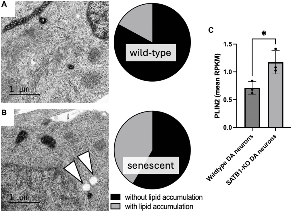
Figure 2.Quantification of lipid accumulation and PLIN2 expression in human stem cell derived DA neurons. Representative TEM images of a mature wild-type (A) and SATB1-KO (senescent) DA neurons (B). Pie charts show the quantification of cells containing LD-like structures. White arrowheads indicate LD-like structures. N = 3, n = 29 per genotype. Scale bar = 1 um. (C) PLIN2 expression in wildtype and SATB1-KO (senescent) DA neurons. Student’s t-test was performed. *p < 0.05.