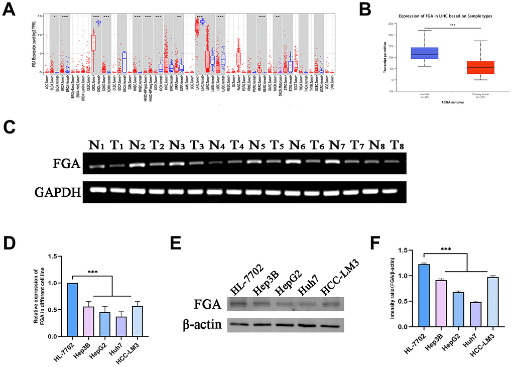
Figure 1.FGA is underexpressed in liver cancer tissue. (A) Analysis of differential FGA expression between liver cancer and adjacent non-cancerous tissues using TCGA RNA-seq data through the TIMER database. (B) Evaluation of FGA expression levels in hepatocellular carcinoma using UNCLAN data. (C) Detection of FGA expression in eight cases of adjacent non-cancerous tissue and eight cases of liver cancer tissue through agarose gel electrophoresis assay. (D) Detection of FGA mRNA expression in normal liver cell line HL-7702 and different liver cancer cell lines using RT-qPCR. (E) Detection of FGA protein expression in normal liver cell line HL-7702 and different liver cancer cell lines using Western blot, and its quantitative analysis. (F) (N: normal tissue, T: tumor tissue; two-tailed Student’s t test; mean ± SD, n = 3; *P < 0.05, **P < 0.01, ***P < 0.001, compared to the normal group).