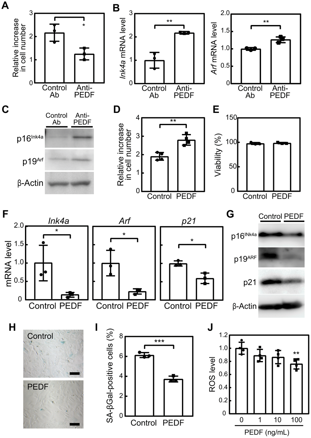
Figure 2.PEDF mediates the anti-cellular senescence effects of C2C12-CM. (A–C) MEFs were cultured in the presence of C2C12-CM treated with a control or PEDF antibody for 3 days. (A) Cell numbers were counted and relative changes in cell numbers in 3 days were plotted. (B) Total RNA was isolated from MEFs and the expression of Ink4a and Arf was analyzed by real-time PCR. Values were normalized to Gapdh in each sample. (C) p16INK4a and p19ARF levels were analyzed by immunoblotting. β-Actin was used as the loading control. (D) MEFs were cultured in the presence of a recombinant of PEDF (100 ng/mL) for 3 days. Changes in cell numbers were plotted. (E) Cell viability was determined by the trypan blue exclusion assay. (F) The expression of Ink4a, Arf, and p21 was analyzed by real-time PCR. Values were normalized to Gapdh in each sample. (G) p16INK4a, p19ARF, and p21 levels were analyzed by immunoblotting. β-Actin was used as a loading control. (H) Cells were stained for SA-β-gal. Scale bar, 100 μm. (I) The percentage of SA-β-gal-positive cells was plotted. (J) Cells were stimulated with the indicated concentrations of recombinant PEDF for 3 days. Intracellular ROS levels were analyzed in each sample, and relative values were plotted against the average of the control sample. Values represent means ± SD. Data were analyzed by the Student’s t-test (A, B, D–F, I) or a one-way ANOVA and Tukey’s post-hoc analysis (J). *P <0.05, **P <0.01, and ***P <0.001.