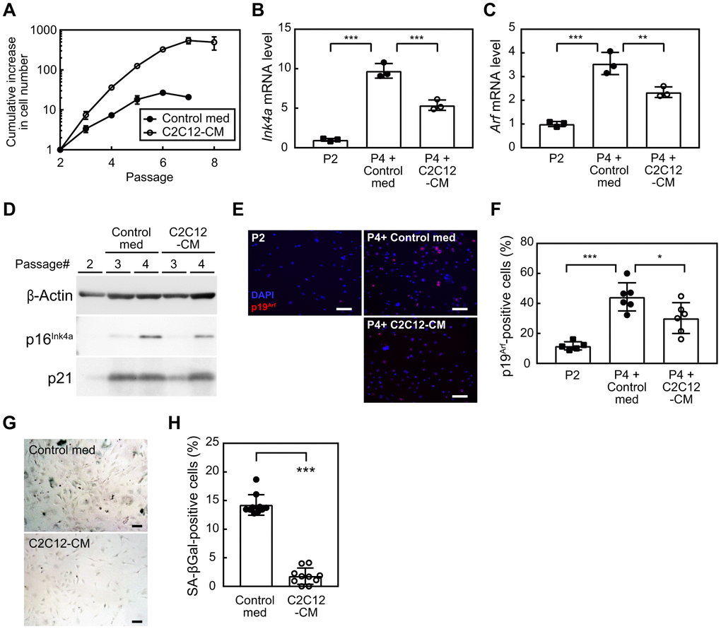
Figure 1.Myocyte-derived factors suppress cellular senescence. (A) Wild-type MEFs were cultured according to the 3T3 protocol in the presence of a control medium or C2C12-CM (1:1 dilution with DMEM containing 10% serum). Data represent the average value of triplicate samples ± SD. (B, C) Total RNA was isolated from cells and the expression of Ink4a (B) or Arf (C) was analyzed by real-time PCR. Values were normalized to Gapdh in each sample. (D) The expression of p16INK4a and p21 was analyzed by immunoblotting. β-Actin was used as a loading control. (E) An immunofluorescence analysis was performed using the p19ARF antibody. Cells were counterstained with DAPI. Scale bar, 100 μm. (F) The number of p19ARF-positive cells was counted in (E). (G) Cells (passage 4) were stained for SA-β-gal. Scale bar, 100 μm. (H) The number of SA-β-gal-positive cells was counted in each sample. Scale bar, 100 μm. Values represent means ± SD. Data were analyzed by a one-way ANOVA and Tukey’s post-hoc analysis (B, C, F) or the Student’s t-test (H). *P <0.05, **P <0.01, and ***P <0.001.