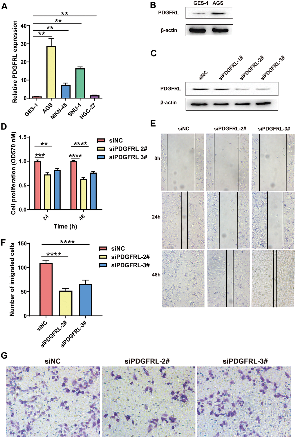
Figure 6.Functional validation for PDGFRL in GC cells. (A) PDGFRL mRNA expressions in normal gastric cells GES-1 and GC cells AGS, MKN-45, SUN-1 and HGC-27. (B) PDGFRL protein expressions in GES-1 and AGS cells. (C) AGS cells were transiently transfected with three different PDGFRL siRNAs (siPDGFRL-1#, siPDGFRL-2# and siPDGFRL-3#) and their negative control (siNC). Western blot was used to measure PDGFRL expressions. β-actin was used as loading control. MTT assay (D), wound healing assay (E) and transwell assay (F) were used to measure proliferation, migration and invasion of AGS cells, respectively. Representative images of the transwell assay were shown (G). The data were presented as the mean ± standard deviation. *** P < 0.01, *** P < 0.001 and **** P < 0.0001.