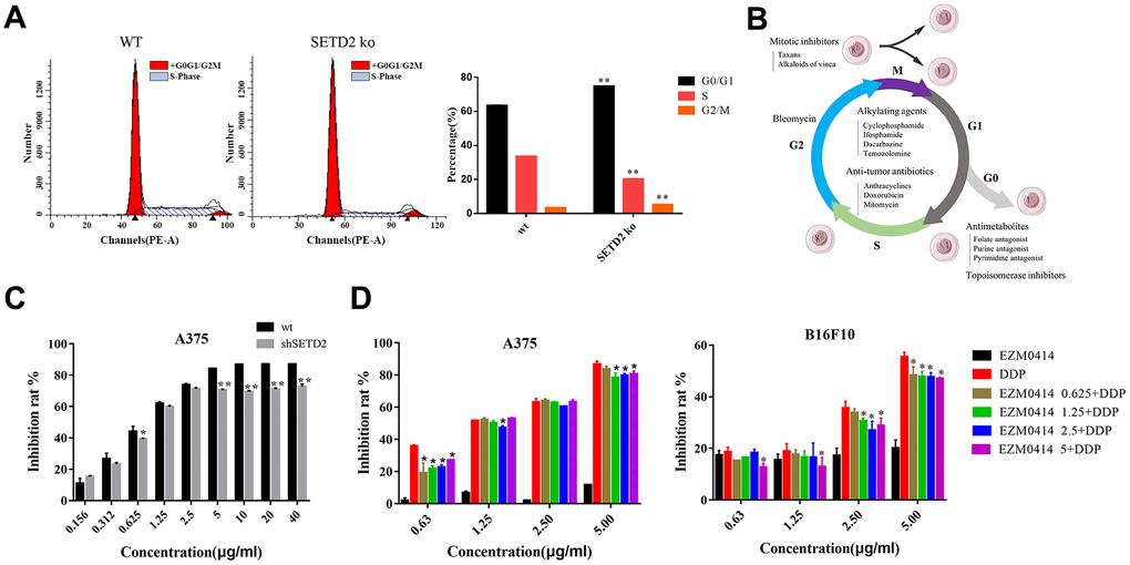
Figure 3.The impact of SETD2 dysfunction on the proliferation inhibition of DDP in melanoma cell lines in vitro. (A) The cell cycle of SETD2 ko A375 cells compared with its wild-type counterparts by flow cytometry. (B) The classification of chemotherapeutics. (C) The proliferation inhibition of SETD2 ko A375 cells cultured in DDP at 0.156, 0.312, 0.625, 1.25, 2.5, 5, 10, 20, 40 μg/ml. (D) CCK-8 testing the proliferation inhibition of A375 cells (left) and B16F10 cells (right) cultured in EZM0414 at 0.625, 1.25, 2.5, 5 μg/ml, DDP at 0.625, 1.25, 2.5, 5 μg/ml, then single or combination drugs were used for 72 hours.