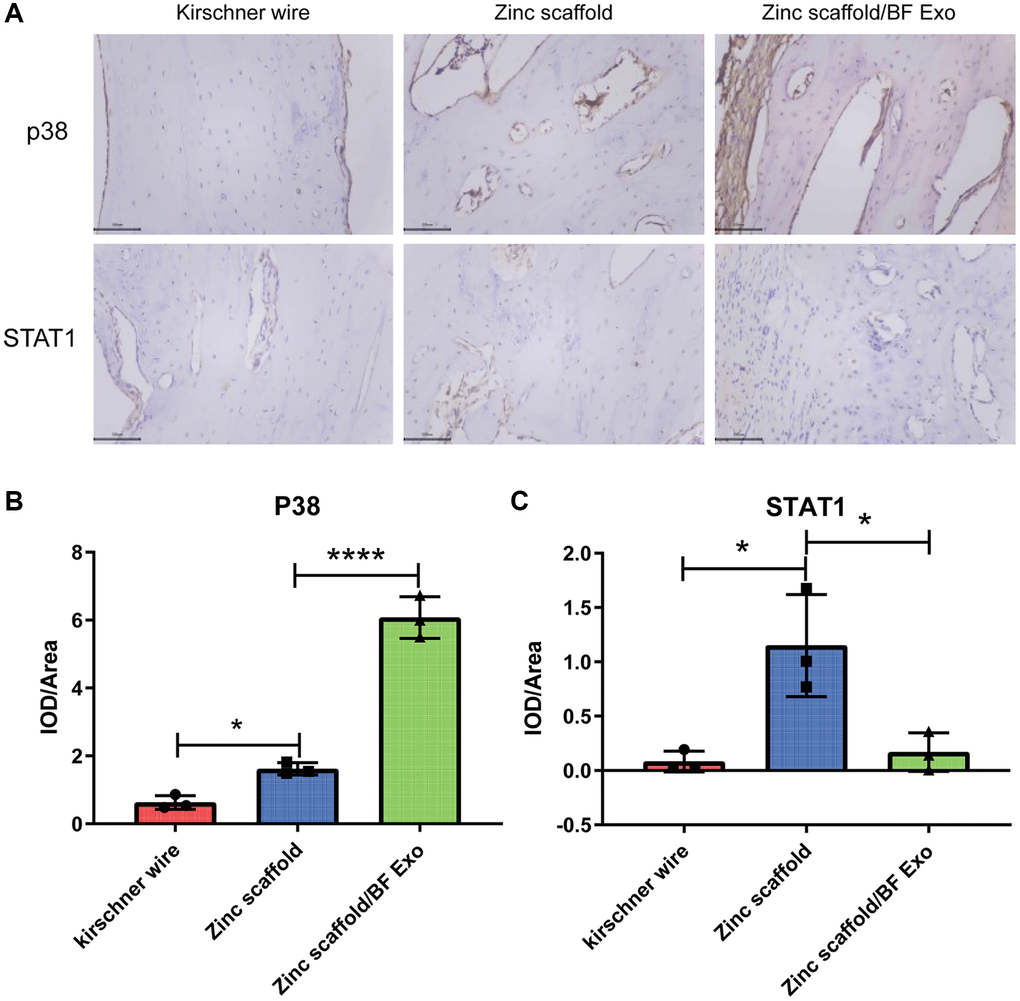
Figure 9.Immunohistochemical analysis of in vivo implantation of zinc scaffold/BF Exo composite implant. (A) Immunohistochemical staining results of rabbit radius in each group after 12 weeks of modeling; (B) Quantitative analysis results of p38 in rabbit radius in each group after 12 weeks of modeling; (C) Quantitative analysis results of STAT1 in rabbit radius in each group after 12 weeks of modeling (*P < 0.05, ****P < 0.0001, nsP > 0.05 vs. Zinc scaffold, n = 3).