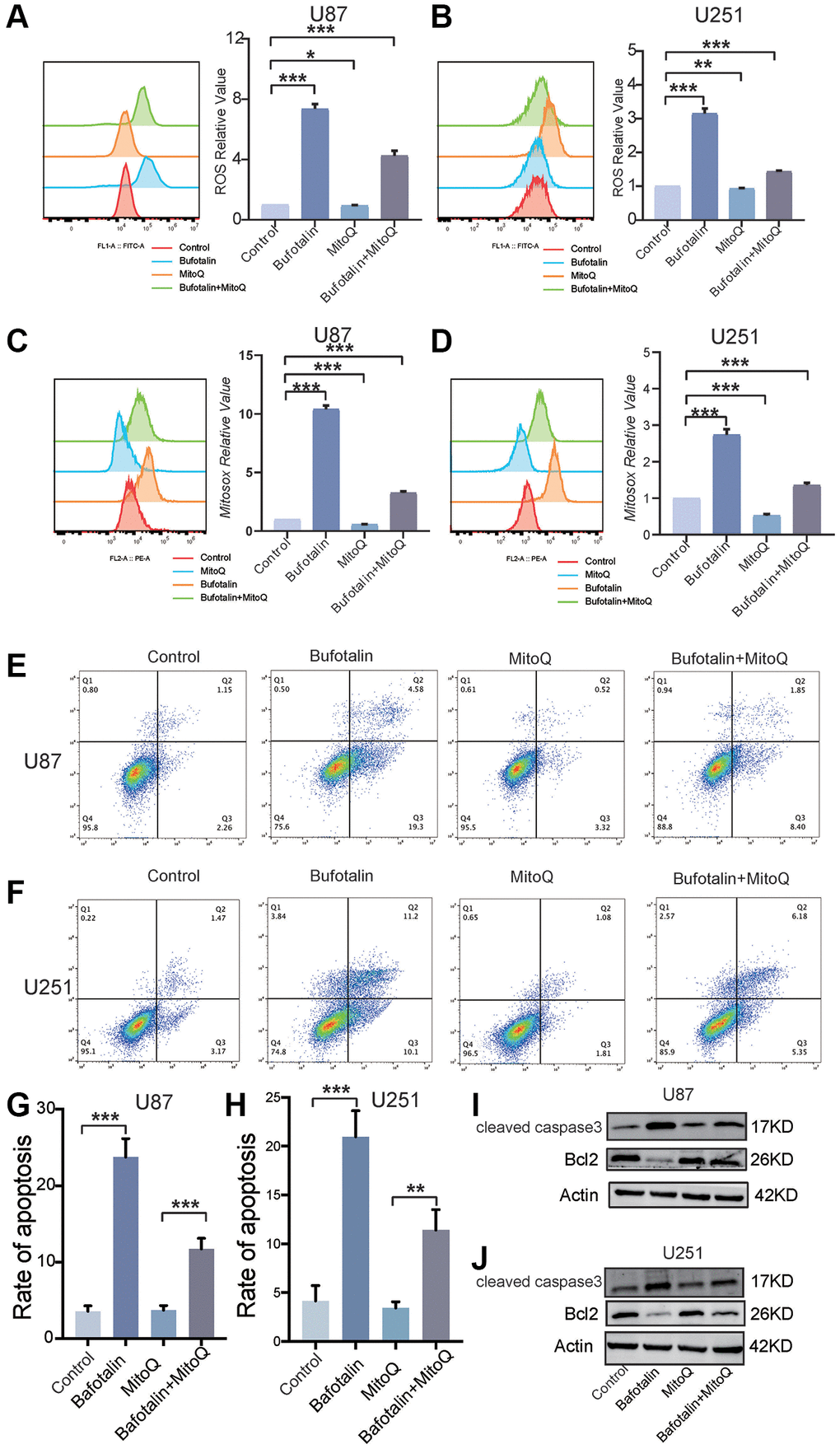
Figure 6.Protecting mitochondria decreased the effect of bufotalin in vitro. (A, B) ROS quantification of cells treated with MitoQ and bufotalin was determined by flow cytometry using the H2DCFDA. (C, D) mitoROS quantification of cells treated with MitoQ and bufotalin was determined by flow cytometry using the MitoSox. (E–H) Cell apoptosis was measured by flow cytometry. U87 and U251 cells were treated with MitoQ and bufotalin for 24 h. (I, J) The expression levels of Bcl2, cleaved caspase-3 and p-AKT were analyzed by western blot. Data are the mean ± SD of triplicate samples. Significant differences compared with the control are indicated by *p < 0.05, **p < 0.01, and ***p < 0.001.