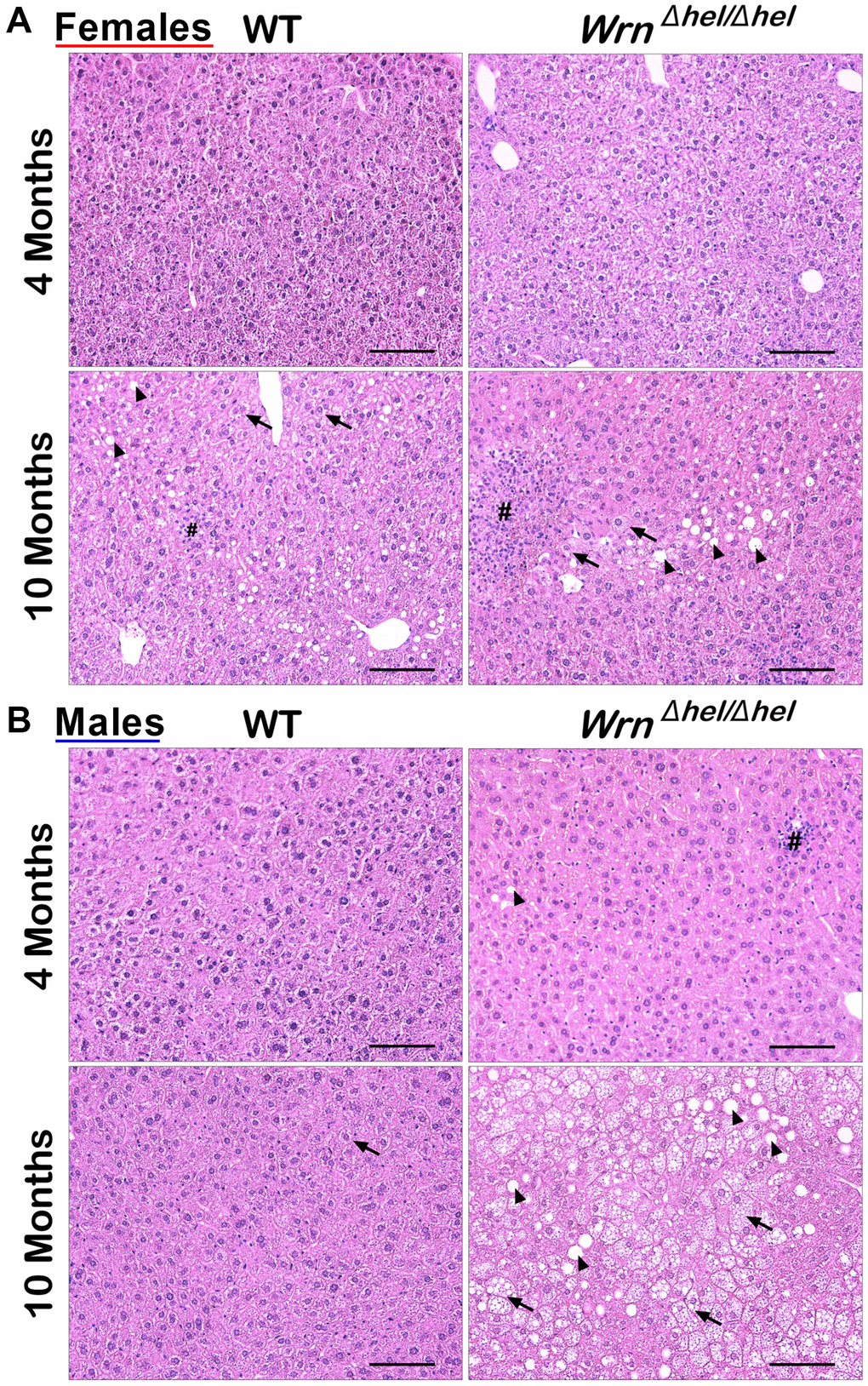
Figure 2.Representative histology of the liver in wild type and WrnΔhel/Δhel mice at four and ten months of age. (A) Representative histological sections from females. (B) Representative histological sections from males. All photomicrographs: hematoxylin and eosin; magnification 200X. Scale bars on the images represent 100 μm. # = inflammatory cell aggregation; arrowheads = macrovesicles; arrows = cytoplasmic microvesicles.