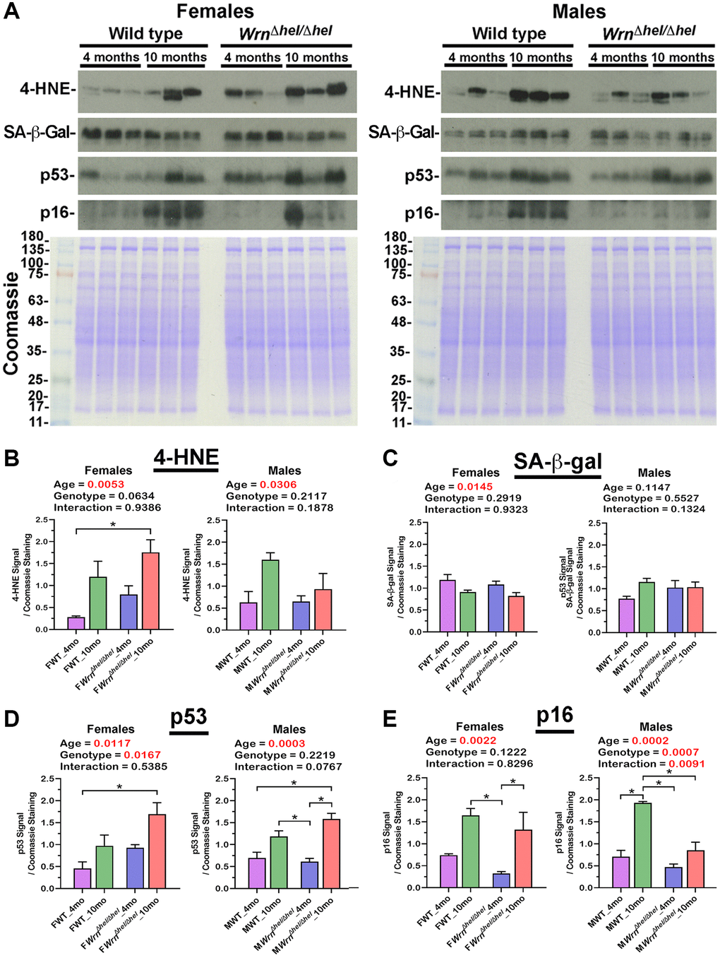
Figure 12.Immunoblot analyses of markers of lipid peroxidation and senescence in the liver of wild type and WrnΔhel/Δhel mice. (A) Examples of western blots showing the levels of 4-hydroxynonenal (4-HNE)-protein adducts, senescence-associated ß-galactosidase (SA-ß-gal), p53, and p16 proteins in both female and male groups. The lower panels show Coomassie staining of the gels. The numbers on the left of the Coomassie represent the molecular weights in kDa. (B) Ratio of the 4-HNE signal over Coomassie staining from the western blots. (C) Ratio of the SA-ß-gal signal over Coomassie staining from the western blots. (D) Ratio of the p53 signal over Coomassie staining from the western blots. (E) Ratio of the p16 signal over Coomassie staining from the western blots. All the graphs represent the mean of each group. Bars represent the SEM. Two-way ANOVA p-values for age, genotype, and the interaction (age x genotype) are indicated on top of each graph. Two-way ANOVA followed by Tukey’s multiple comparisons test p-value < 0.05 are indicated by * in the graphs for each comparison. (N = 3 males or females per group).