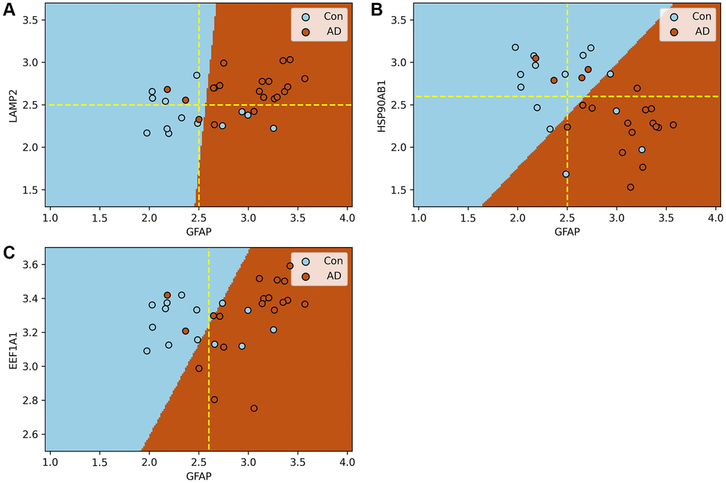
Figure 7.Support vector machine models of the process of substrate translocation into lysosomes during CMA. (A–C) plot the expression of GFAP against that of other key CMA proteins. Here, blue and brown markers represent control and AD groups, respectively, with dashed lines indicating critical expression thresholds. Subfigure A demonstrates that when both LAMP2A and GFAP expressions surpass their thresholds, the risk of AD nears certainty. Subfigure B shows a heightened AD risk when HSP90AB1 falls below its threshold, while GFAP’s expression is above its own. Collectively, these models confirm the sensitivity of the process of substrate translocation into lysosomes during CMA to the progression of AD, highlighting its potential as a biomarker. The molecular rationale underlying these observations involves CMA’s activation in response to the excessive accumulation of abnormal proteins due to AD progression, necessitating a three-step process for substrate degradation. Initially, HSP90AB1 facilitates substrate unfolding to prepare for lysosomal delivery. Subsequently, LAMP2A and GFAP collaborate to form a translocation complex, efficiently directing substrates to the lysosome. Finally, EEF1A1 disengages GFAP from the complex, resetting LAMP2A for subsequent cycles. These stages correspond to the findings depicted in Subfigures B, A, and C, respectively. Subfigure B underscores the initial response of the substrate translocation process into lysosomes within CMA to proteotoxicity accumulation, a critical factor in AD risk assessment. Subfigure A showcases the delivery phase, where the combined actions of LAMP2A and GFAP, manifested through their increased expression levels, significantly boost the process’s capacity to eliminate proteotoxic accumulations. This stage indicates the proactive engagement of this specific CMA phase in substrate degradation. Thus, the integrated function of this lysosomal entry process, rather than the action of individual proteins, stands out as a prominent biomarker for AD. A more comprehensive explanation of this process and its implications for AD diagnosis is provided in Conclusion and illustrated in Figure 6.