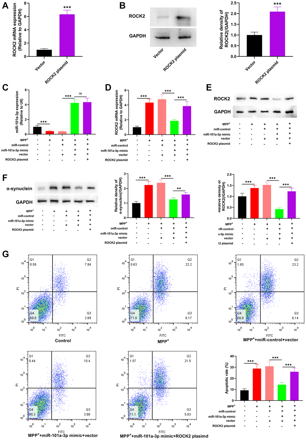
Figure 6.miR-101a-3p reduces the damage of Neuro-2a cells induced by MPP+ via inhibiting ROCK2. To confirm the mechanisms of miR-101a-3p and ROCK2 in MPP+-induced injury of Neuro-2a cells, miR-101a-3p mimics and ROCK2 overexpression plasmids were transfected into Neuro-2a cells before Neuro-2a cells were exposed to MPP+. There were 5 groups: control group, MPTP group, MPP++miR-control+vector group, MPP++miR-101a-3p mimic+vector group, and MPP++miR-101a-3p mimic+ROCK2 plasmid group. (A) qRT-PCR was employed to verify the change in ROCK2 mRNA expression after Neuro-2a cells were transfected with ROCK2 overexpression plasmids. (B) Western blot was employed to examine the change in ROCK2 protein expression after Neuro-2a cells were transfected with ROCK2 overexpression plasmids. (C) Detection of miR-101a-3p expression in each group of cells through qRT-PCR. (D) ROCK2 mRNA expression in each group of cells was detected by qRT-PCR. (E) Western blot detection of ROCK2 protein expression level in each group of cells. (F) Western blotting was conducted to detect α-synuclein protein expression in each group of cells. (G) Flow cytometry was conducted to detect the apoptosis level in each group. **P < 0.01 and ***P < 0.001.