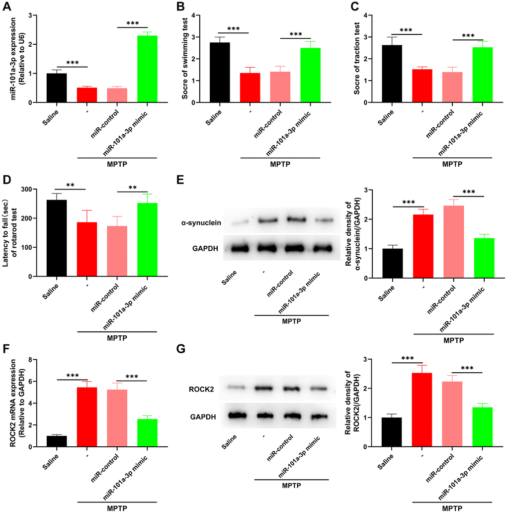
Figure 3.miR-101a-3p mimic inhibits ROCK2 expression and neurological damage in PD mice. To confirm the role of miR-101a-3p in PD mice, miR-101a-3p mimics were intracerebroventricularly injected into the mice to overexpress miR-101a-3p before intraperitoneal injection of MPTP into the mice. There were 4 groups: saline group, MPTP group, MPTP+miR-control group, and MPTP+miR-101a-3p mimic group (n = 3 in each group). (A) qRT-PCR was performed to detect the expression levels of miR-101a-3p in mice in each group. (B) Swimming test was conducted to score the motor ability of each group of mice. (C) Traction test was conducted to score the balance ability of each group of mice. (D) Rotarod test was conducted to score the balance ability of each group of mice. (E) Western blot was performed to detect α-synuclein protein expression in each group of mice. (F) Detection via qRT-PCR of ROCK2 mRNA expression level in each group of mice. (G) Western blot detection of ROCK2 protein expression levels in each group of mice. **P < 0.01 and ***P < 0.001.