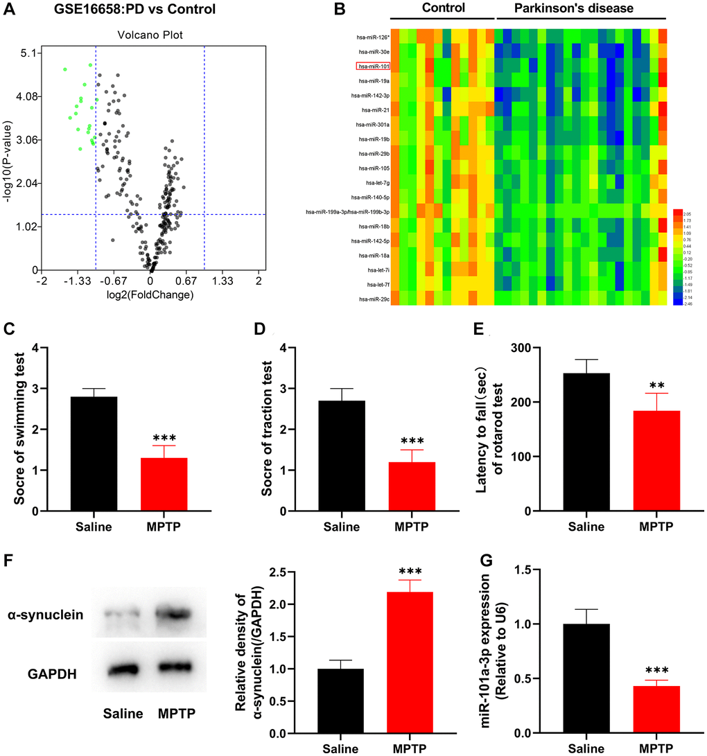
Figure 1.miR-101a-3p is lowly expressed in peripheral blood mononuclear cells of PD patients and brain tissues of mice with PD. (A) The volcano plot shows the differentially expressed miRNAs in the in peripheral blood mononuclear cells of PD patients (vs. healthy controls) in the GSE16658 dataset. (B) The heat map shows the expression profile of significantly down-regulated miRNAs in GASE16658. (C) Swimming test was adopted to score the motor ability of mice in normal saline group and MPTP group (n = 3 in both groups). (D) Traction test was adopted to score the balance ability of mice in normal saline group and MPTP group (n = 3 in both groups). (E) Rotarod test was adopted to evaluate the balance ability of mice in normal saline group and MPTP group (n = 3 in both groups). (F) Western blotting was applied to detect the expression of α-synuclein protein in mice of the normal saline group and MPTP group (n = 3 in both groups). (G) qRT-PCR was applied to detect miR-101a-3p expression in SNpc of mice in the normal saline group and MPTP group (n = 3 in both groups). **P < 0.01 and ***P < 0.001.