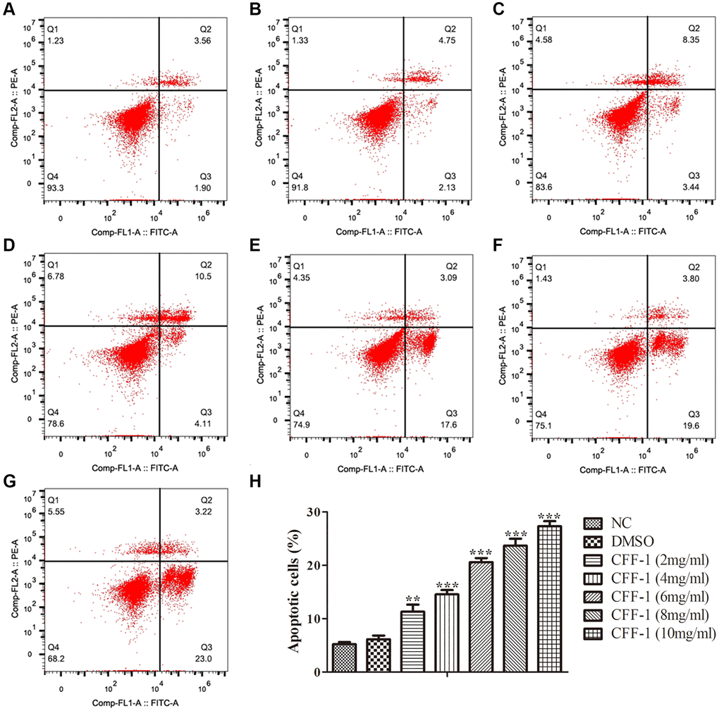
Figure 7.Apoptosis analysis of CFF-1-treated PC-3 cells by flow cytometry after 24 hours of treatment. Each panel corresponds to a different treatment condition: (A) NC, (B) DMSO, and (C–G) increasing concentrations of CFF-1 at 2, 4, 6, 8, and 10 mg/ml, respectively. (H) Quantitative analysis of the percentage of apoptotic cells across different treatment conditions. **p < 0.01, ***p < 0.001.