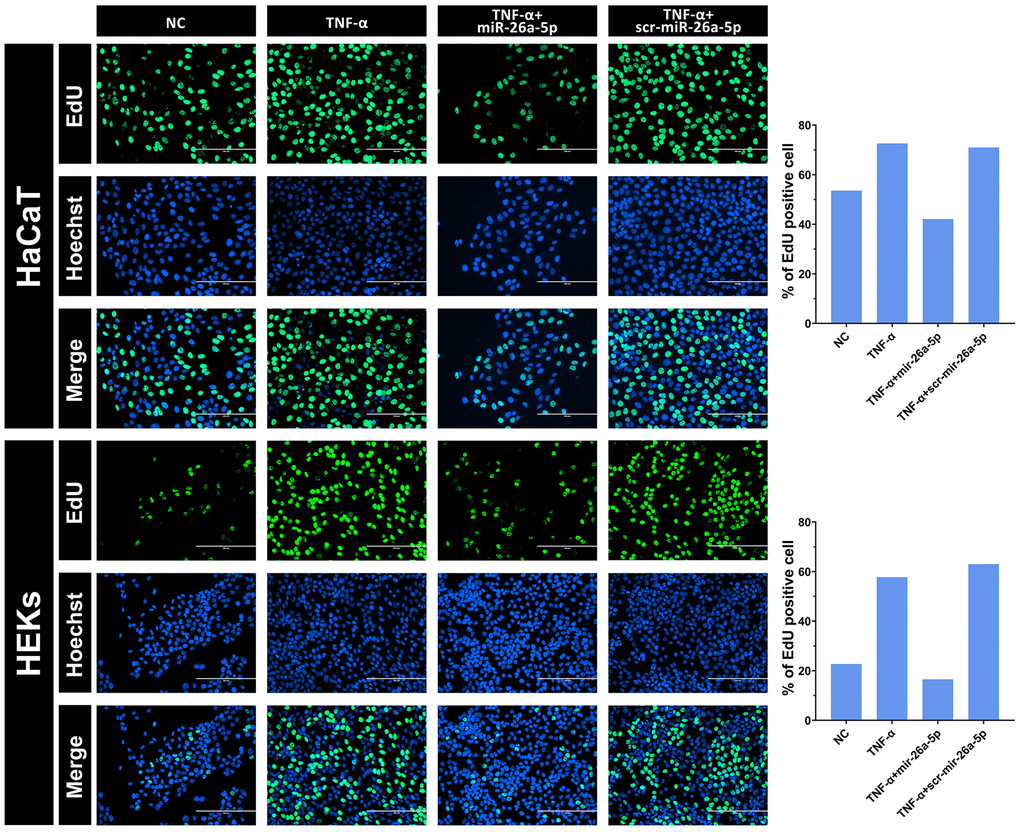
Figure 4.Intuitive observation of the cell proliferation rate of HaCaT keratinocytes and HEKs by EdU staining. After 48 hours, the cells were transfected with 50 nM miRNA mimics. The percentage of EdU incorporation was analyzed using Image-Pro Plus 6.0 software. Scale bars: 200 μm.