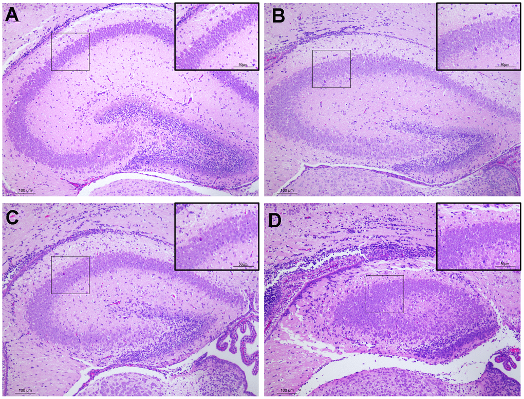
Figure 2.Histopathological changes in the hippocampus tissues of male offspring mice following pregnancy exposure to DEPs through hematoxylin and eosin (H&E) staining. 100 or 200 × magnification. (A) Control group. The morphology of hippocampus tissues was normal. (B–D) DEPs (0.235, 0.47 and 0.94 μg/mouse) treatment groups. Scale bar: 100 μm. N=4.