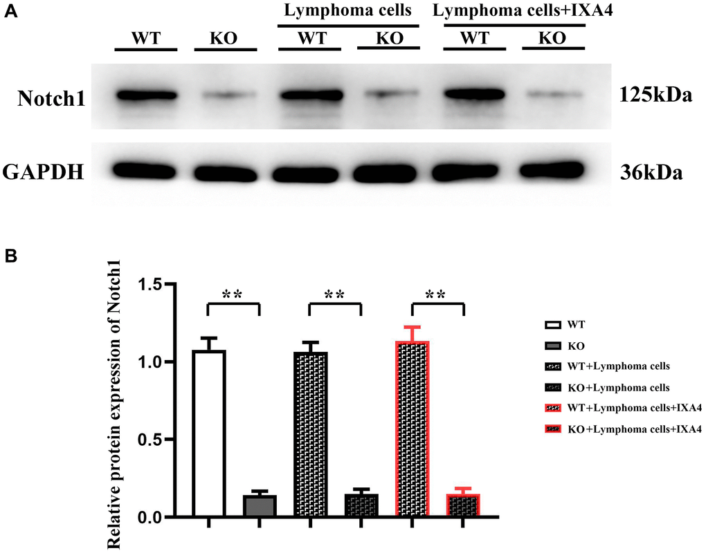
Figure 1.Western blotting of Notch-1. (A) Protein bands of Notch-1; (B) Relative protein expression of Notch-1. (WT group vs. KO group; WT + lymphoma cell group vs. KO + lymphoma cell group; WT + lymphoma cell + IXA4 group vs. KO + lymphoma cell + IXA4 group; **P < 0.01, nsP > 0.05; N = 3).