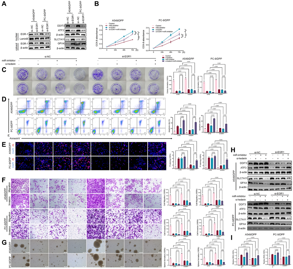
Figure 8.α-Hederin promoted EGR1 nuclear translocation and directly repressed miR-96-5p in NSCLC. (A) Western blot demonstrating EGR1 protein levels in nuclear and cytosol. DDIT3, ATF3, GPX4, and SLC7A11 protein levels are shown following transfection with si-EGR1 for 24 hours. (B) CCK8 results showing the viability of NSCLC cells after transfection with si-EGR1 or miR-96-5p mimic for 24 hours. (C) Colony formation assay showing proliferation in NSCLC cells after treatment with α-Hederin (50 μM) for 24 hours and transfection with si-EGR1 or miR-96-5p mimic for 24 hours. (D) Cell apoptosis was detected by flow cytometry. (E) Propidium iodide (PI) staining (red) indicates apoptotic/necrotic cells. (F) Cell migration and invasion were detected by Transwell assay. (G) Spheroid formation ability assessed in NSCLC cells. (H) Western blot demonstrating EGR1, DDIT3, ATF3, GPX4, and SLC7A11 protein levels in nuclear and cytosol of NSCLC cells. (I) Cellular ROS levels were detected in NSCLC cells after treatment with α-Hederin (50 μM) for 24 hours and transfection with si-EGR1 or miR-96-5p mimic for 24 hours. (n = 3). Data are shown as mean ± SD, One-way ANOVA, *P < 0.05, **P < 0.01, ***P < 0.001.