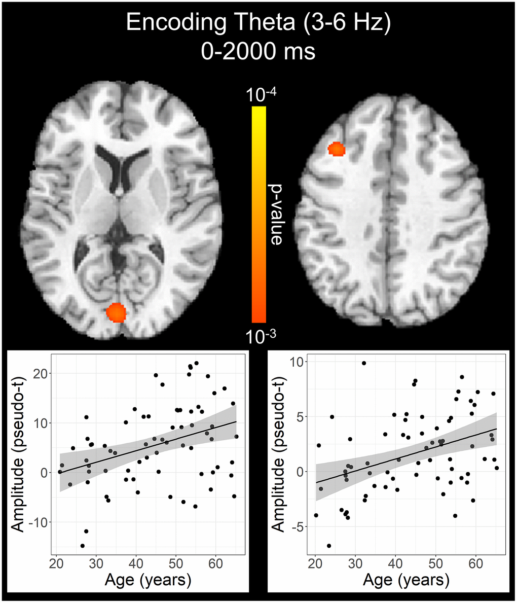
Figure 4.Effect of age on theta oscillations during the encoding phase. Whole-brain linear regression analysis revealed that theta activity increased in the left primary visual cortex and left dlPFC with older age. Linear regression plots of peak voxel pseudo-t values are shown as a function of age. Lines of best-fit and 95% CI (shaded area) are overlaid.