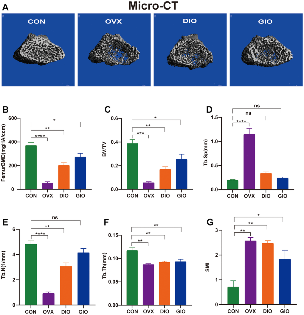
Figure 1.(A) Representative 3D micro-CT reconstructions of femurs from per group. (B–G) Trabecular bone at distal femoral metaphysis after 10 weeks. Parameters included BMD, BV/TV, Tb.Sp, Tb.N, Tb.Th, and SMI. Data are means ± standard error of the mean (SEM). n = 6, *P < 0.05, **P < 0.01, ***P < 0.001, and ****P < 0.0001; ns, no significance.