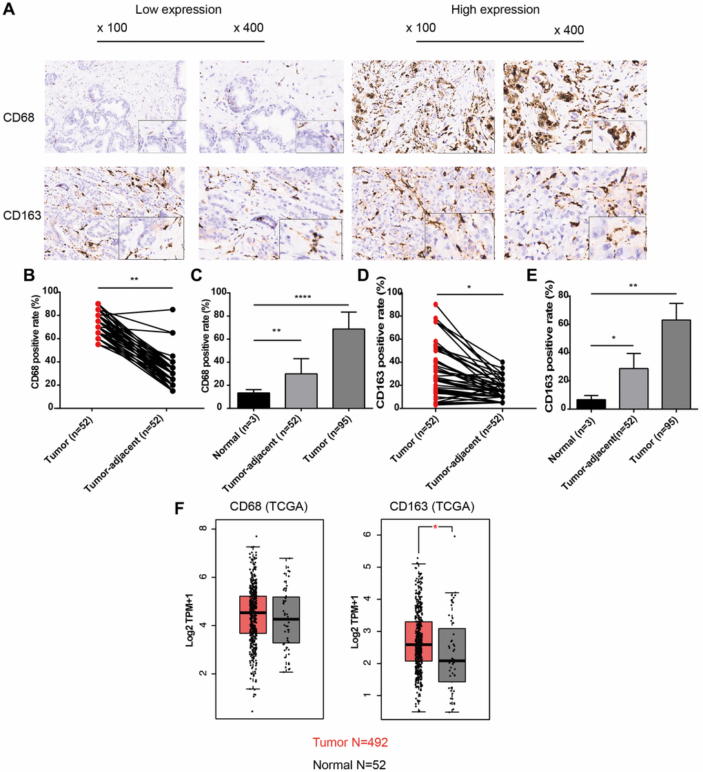
Figure 1.Expression patterns of CD68 and CD163 proteins in PCa and normal prostate tissue. (A) Representative IHC images of PCa tissue slides with low (left panel) or high (right panel) levels of CD68 and CD163 protein. IHC, immunocytochemistry. (B) Dot plot shows the positive rate of CD68 in PCa and para-cancer (para-PCa) tissue. (C) Bar plot shows the positive rate of CD68 in normal prostate tissue, para-PCa, and PCa. (D) Dot plot shows the positive rate of CD163 in PCa and para-PCa tissue. (E) Bar plot shows the positive rate of CD163 in normal prostate tissue, para-PCa, and PCa. (F) Box plots showed the mRNA expression levels of CD68 and CD163 in the TCGA-PRAD dataset. Student’s t-test, *P < 0.05, **P < 0.01, ****P < 0.001.