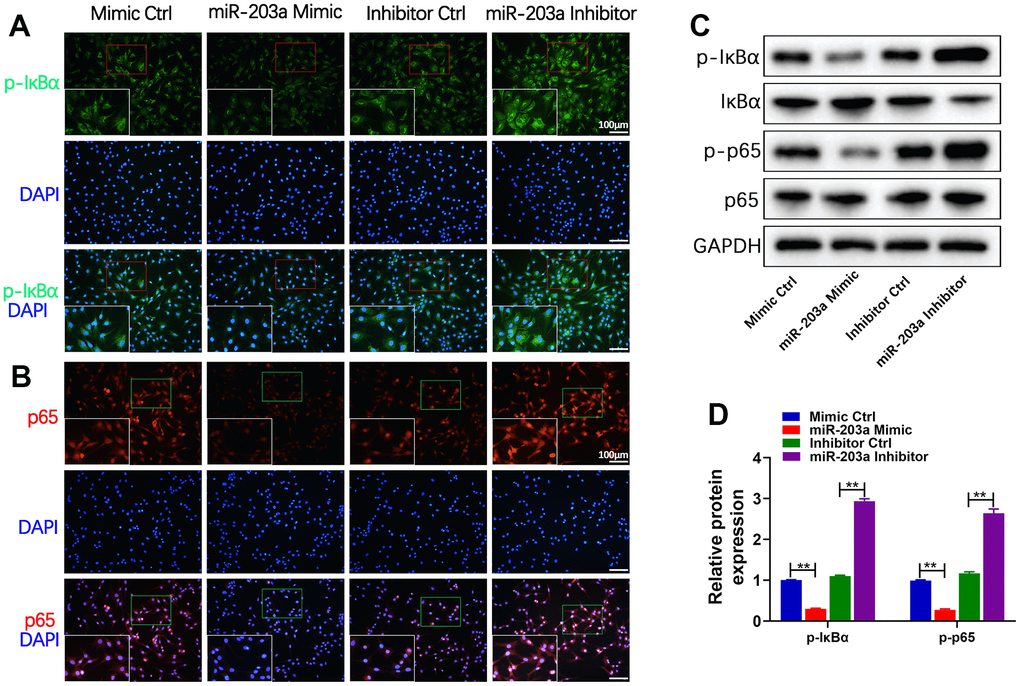
Figure 5.miR-203a-3p inhibited the NF-κB signaling pathway in chondrocytes. (A, B) Immunofluorescence staining showing the phosphorylation of IκBα (green) and the nuclear translocation of p65 (red) in LPS-induced chondrocytes following transfection with miR-203a-3p mimic, inhibitor and consistent negative controls. (C, D) WB analysis and the relative quantification showing the expression levels of proteins related with NF-κB signaling pathway (p-IκBα, IκBα, p-p65 and p65). All experiments were performed in triplicated and data were presented as the mean±SD, n=3 per group. *P<0.05, **P<0.01.