Submit an Article
Navigate
Home
Editorial Board
Editorial Policies
Current Volume
Archive
Scientific Integrity
Publication Ethics Statements
Interviews with Outstanding Authors
Newsroom
Sponsored Conferences
Podcast
Contact
Special Collections
Submit an Article
Online ISSN: 1945-4589
Research Paper
|
Volume 15, Issue 24
|
pp. 14945–14956
Imaging of brain clearance pathways via MRI assessment of the glymphatic system
Back to article
Figure 5
(5 of 6)
−
100%
+
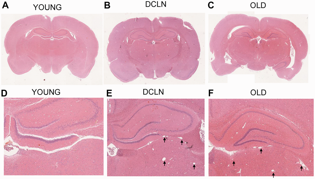
Figure 5.
HE-staining histology results from YOUNG (
A
,
D
), DCLN (
B
,
E
), and OLD (
C
,
F
) SD rats. The black arrows indicate the dilated perivascular spaces in the brain.