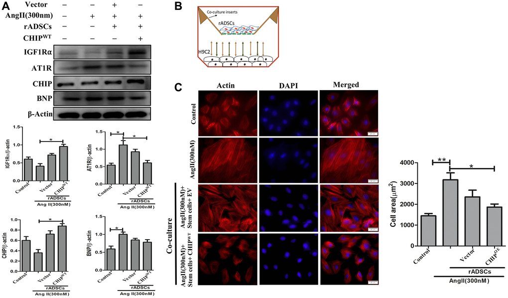
Figure 5.CHIP-overexpressing ADSCs attenuate Ang II-induced hypertrophy in H9c2 cells. (A) The co-culture analysis with rADSCs and H9c2 to determine the expression levels of survival and hypertrophic markers in H9c2 cells. (B) The schematic diagram of the co-culture technique showing rADSCs in the upper chamber and H9c2 cells in the bottom chamber. (C) Rhodamine-phalloidin staining measures the area of H9c2 cells after being challenged with Ang II for 24 h. Scale bar = 20 μm. (N = 3; *p < 0.05; **p < 0.01 indicate significant differences).