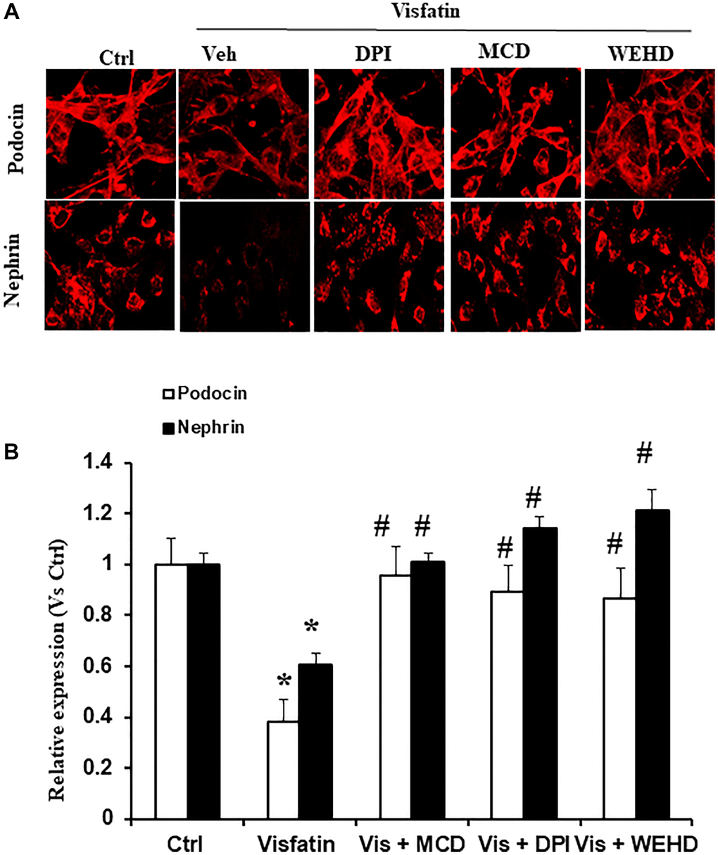
Figure 4.Effects of Visfatin on podocyte injury. (A) Representative immunofluorescence staining of podocin, nephrin, in podocytes with or without stimulation of visfatin, DPI or MCD or WEHD (original magnification, 400x). (B) Summarized data show the percentage of podocyte positive for podocin and nephrin (n = 6). Values are means ± SEM, showing fold changes as compared with control. Abbreviations: Ctrl: Control; Veh: Vehicle; Vis: Visfatin; DPI: diphenyleneiodonium; MCD: methyl-β-cyclodextrin. *P < 0.05 vs. control; #P < 0.05 vs. visfatin.