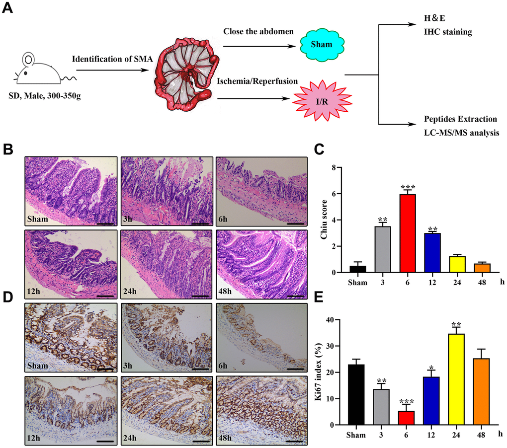
Figure 1.Establishment of the rat IIRI model. (A) Schematic diagram of the experimental design. (B–E) Representative images of intestinal sections from rats of the sham operated and I/R groups stained with H&E (B) or the IHC marker Ki67 (C), respectively, and quantified histopathologically based on Chiu’s score (D) or immunoreactive scores (E), respectively. *P < 0.05; **P < 0.01; ***P < 0.001.