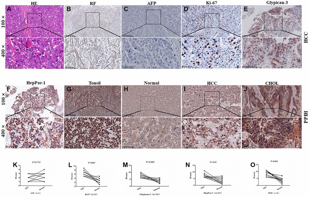
Figure 3.Diagnosis and IHC identification of HCC. (A) HE staining image of HCC. (B) Reticular fiber (RF) staining picture of HCC. (C–F) IHC for AFP, Ki-67, Glypican-3, and HepPar-1 in HCC. (G–J) Representative IHC images of PPIH in tonsil (positive control), HCC, CHOL (cholangiocarcinoma) specimens and matched adjacent normal tissues (H). Regions in squares are magnified 4×; in bottom panels. (K–O) Relative AFP (K) Ki-67 (L) Glypican-3 (M) HepPar-1 (N) PPIH (O) expression scores in HCC and paired normal tissues.