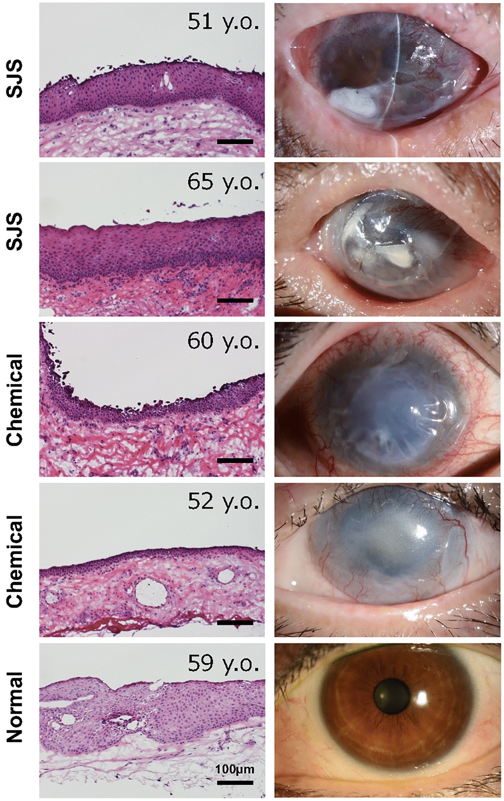
Figure 4.Histology of conjunctival tissues from patients with limbal stem cell deficiency. Representative ocular images of two cases with Stevens-Johnson syndrome (SJS), two cases with chemical injury, and one healthy subject. The histological sections were stained with hematoxylin and eosin stain. Scale bars indicate 100 μm.