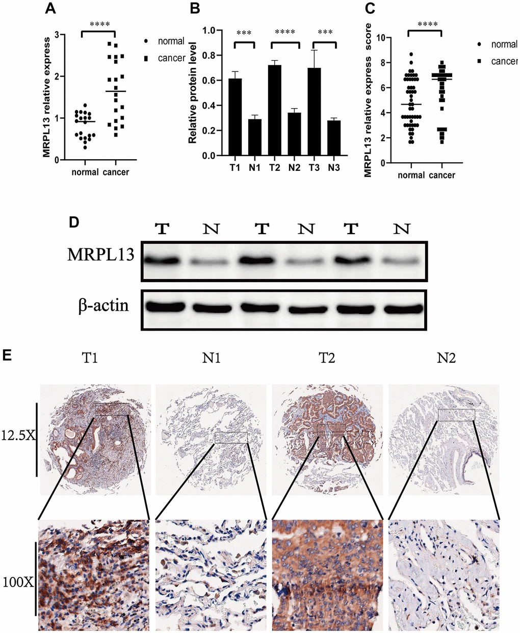
Figure 12.Expression of MRPL13 in cancer and normal tissues. (A) Fluorescence real-time quantitative PCR was used to detect the expression of MRPL13 in tumor tissues and normal tissues adjacent to cancer. (B) Western blot to verify the expression of MRPL13 in cancer and normal tissues adjacent to cancer (the experiment was repeated thrice). (C) Immunohistochemistry score to verify the expression of MRPL13 in cancer and normal tissues adjacent to cancer. (D) Western blot representative picture. (E) Immunohistochemical representative pictures of cancer patients and normal patients (panorama above, detail below). *p-value < 0.05, **p-value < 0.01, ***p-value < 0.001, and ****p-value < 0.0001.