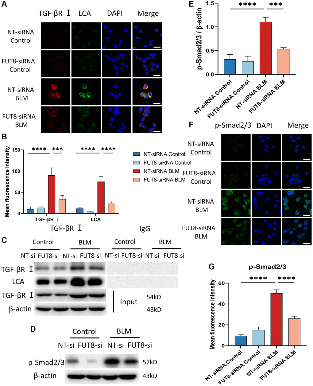
Figure 4.Blocking CF modification of TGF-βR I alleviates activation of TGF-β signaling pathway in BLM-induced AECs senescence. (A, B) Representative immunofluorescence shows the colocalization of TGF-βR I (Red) and LCA (Green) in different treatments with MLE12 cells. DAPI was used to counterstain the nuclei. Scale bar, 25 μm. Data were presented as the mean ± SD. ***P < 0.001. ****P < 0.0001. (C) Immunoprecipitation of TGF-βR I with LCA was tested by immunoblotting analysis. (D, E) Western blot using anti-p-Smad2/3 and anti-β-actin antibodies. Densitometric analyses of the bands were adjusted to be equal to β-actin. Data were presented as the mean ± SD. ***P < 0.001. ****P < 0.0001. (F, G) The p-Smad2/3 expression in different groups was measured via the immunofluorescence technique. DAPI was used to counterstain the nuclei. Scale bar 25 μm. Data were presented as the mean ± SD. ****P < 0.0001.