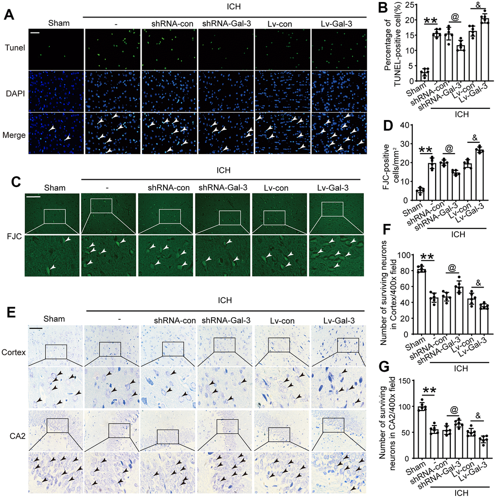
Figure 4.Inhibition of Gal-3 reduced apoptosis and neuron loss induced by ICH. (A, B) Apoptotic cells were labeled in brain sections using TUNEL staining, and the percentage of apoptotic cells was analyzed statistically. Arrow indicated TUNEL positive cells. Nuclei were labeled with DAPI (blue). (C, D) FJC staining. Arrow indicated FJC positive cells. (E–G) Nissl staining was used to assess the loss of neurons in the CA2 region of the hippocampus and in the cortex. Arrow indicated surviving cells. Scale bar = 50um. The black dots represent individual data in each group. **p < 0.01 and *p < 0.05 vs. Sham group, @p < 0.05 vs. ICH+shRNA-con group, &p < 0.05 vs. ICH+Lv-con group, n = 6.