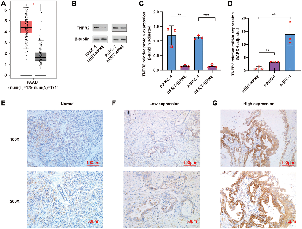
Figure 1.(A) TNFR2 gene was analyzed using GEPIA. (B, C) TNFR2 protein in PANC-1 and ASPC-1 cells were measured by western blot. (D) TNFR2 mRNA in PANC-1 and ASPC-1 cells were detected using qRT-PCR. (E) Immunohistochemical staining in normal pancreatic tissue. (F) Immunohistochemical staining in the low-expression group of pancreatic cancer tissue. (G) Immunohistochemical staining in the high-expression group of pancreatic cancer tissue.