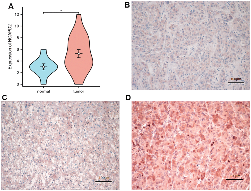
Figure 10.The expression of NCAPD2 protein in normal tissues and LIHC by immunohistochemistry (EnVision; original magnification, ×200). (A) NCAPD2 had higher expression in LIHC than normal tissues. (B) Weak positive expression of NCAPD2 in LIHC. (C) Moderate positive expression of NCAPD2 in LIHC. (D) Strong positive expression of NCAPD2 in LIHC. (*P<0.05).