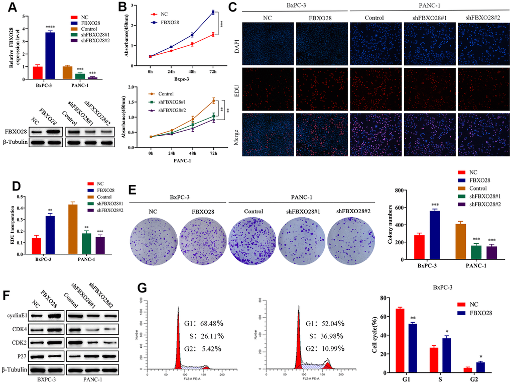
Figure 2.FBXO28 overexpression increases pancreatic cancer cell proliferation. (A) Lentiviral transfection to form stable cells (negative control [NC], FBXO28, Control, shFBXO28#1, shFBXO28#2) and qRT-PCR and western blot to verify transfection effectiveness. (B–E) Cell Counting Kit-8 (CCK-8), EdU, and clone plate experiments were used to identify the capacity of FBXO28 for cell proliferation and formation in pancreatic cancer cells. (F, G). **P < 0.01, ***P < 0.001, ****P < 0.0001.
(H) Western blot and flow cytometry to investigate the effect of FBXO28 on the cell cycle. (I) To construct a xenograft model, mice were injected subcutaneously with cells according to grouping, tumor volume was assessed weekly, the mice were euthanized after 6 weeks, and the tumors were resected and weighed. (J) Immunohistochemical (IHC) of mouse tumor tissues showing the protein expression of Ki67 and proliferating cell nuclear antigen (PCNA) (magnification: ×200, ×400). **P < 0.01, ***P < 0.001, ****P < 0.0001.