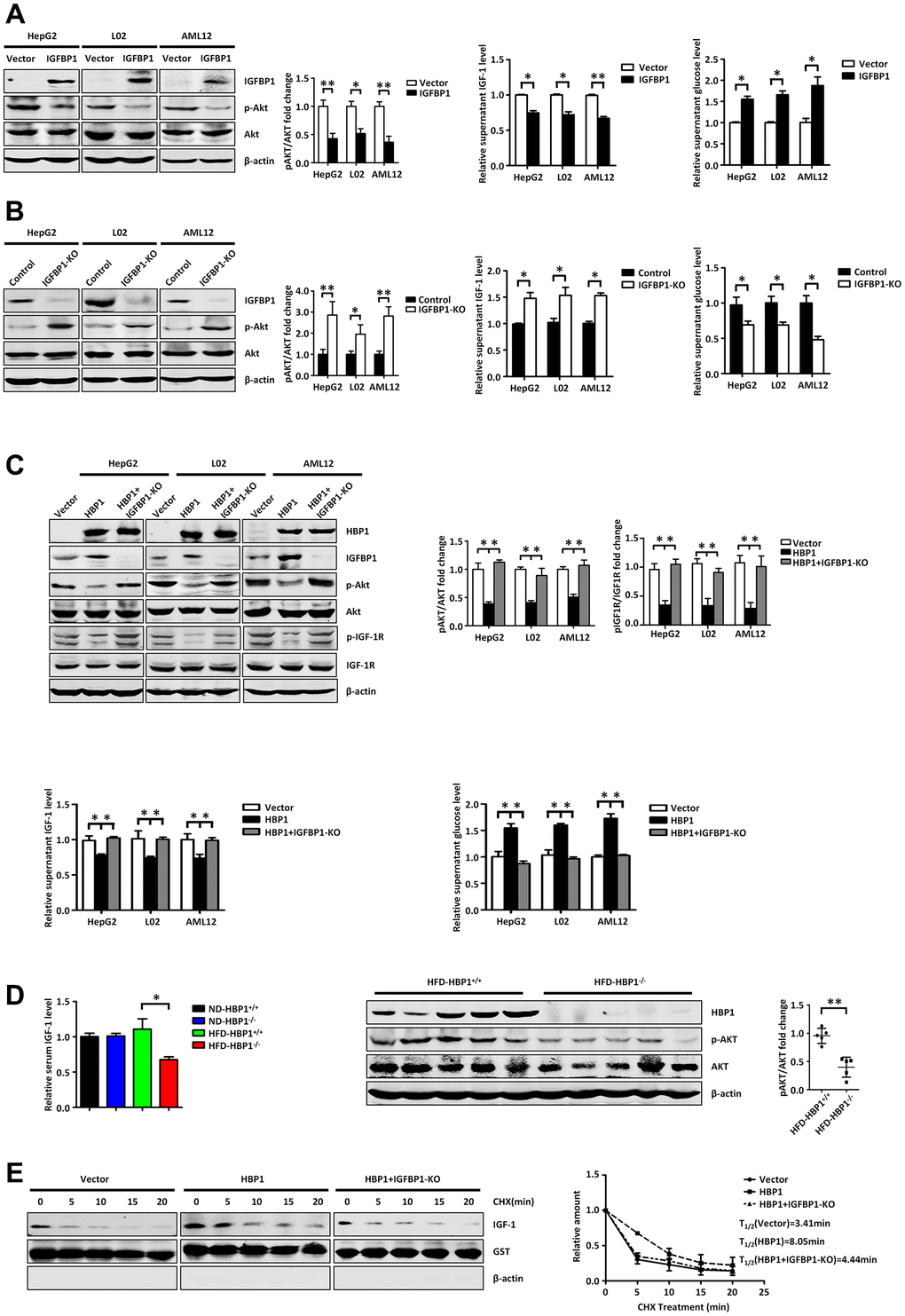
Figure 6.The HBP1-IGFBP1 axis increases extracellular glucose concentration by reducing activation of the PI3K-AKT signaling pathway. (A) IGFBP1 inhibits p-AKT signaling pathway, inhibits supernatant IGF-1 levels and elevates supernatant glucose level. The protein levels of IGFBP1, p-AKT and AKT were measured by western blotting in HepG2, L02 and AML12 cells. β-actin was used as a loading control. The content of IGF-1 in supernatant was measured by ELISA kit. The supernatant was changed from DMEM to EBSS and was collected after 24 hours. Then the supernatant glucose level was measured by glucose testing kit. (B) IGFBP1 knockout activates p-AKT signaling pathway, increases supernatant IGF-1 levels and suppresses supernatant glucose level. The protein levels of IGFBP1, p-AKT and AKT were measured by western blotting in IGFBP1-knockout HepG2, L02 and AML12 cells constructed by CRISPR/Cas 9 system. β-actin was used as a loading control. The content of IGF-1 in supernatant was measured by ELISA kit. The supernatant was changed from DMEM to EBSS and was collected after 24 hours. Then the supernatant glucose level was measured by glucose testing kit. (C) HBP1 can inhibit p-AKT signaling pathway, decreases supernatant IGF-1 levels and elevates supernatant glucose level by IGFBP1. The protein levels of HBP1, IGFBP1, p-AKT, AKT, p- IGF-1R and IGF-1R were measured by western blotting in HepG2, L02 and AML12 cells. β-actin was used as a loading control. The content of IGF-1 in supernatant was measured by ELISA kit. Then the supernatant was changed from DMEM to EBSS and was collected after 24 hours. The supernatant glucose level was measured by glucose testing kit. (D) The level of serum free IGF-1 in HFD-HBP1−/− mice is lower than that of HFD-HBP1+/+ mice (n=5), so that the PI3K-AKT signaling pathway is inhibits. The content of mice serum IGF-1 was measured by ELISA kit. The protein levels of HBP1, p-AKT and AKT were measured by western blotting. β-actin was used as a loading control. (E) HBP1 affects the stability of IGF-1 through IGFBP1. The protein levels of IGF-1, GST and β-actin were measured by western blotting. GST was used as a loading control. Data were the mean ± SD by a two-tail, unpaired Student’s t-test. *, p<0.05. **, p<0.01.