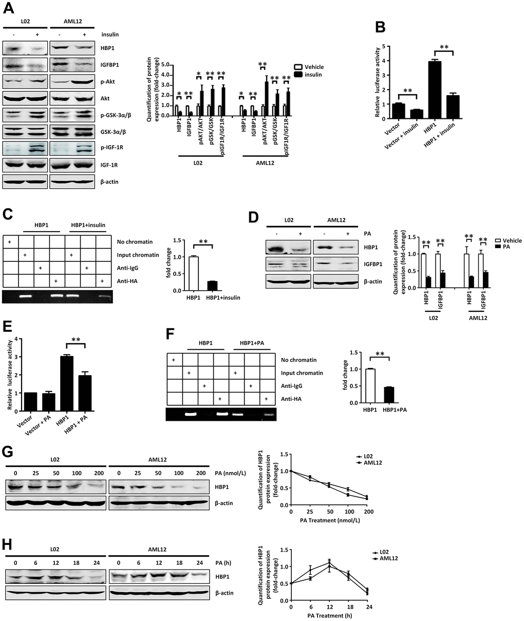
Figure 5.Insulin and palmitic acid suppress the protein expression of HBP1 and the combination of HBP1 and IGFBP1 promoter. (A) Insulin activates p-AKT signaling pathway and inhibits protein expression of HBP1 and IGFBP1. The protein levels of HBP1, IGFBP1, p-AKT, p-GSK-3α/β, p-IGF-1R, AKT, GSK-3α/β and IGF-1R were measured by western blotting in L02 and AML12 cells. β-actin was used as a loading control. Quantification was normalized to β-actin. (B) Insulin can attenuate both the IGFBP1 promoter activity and the activation effect of HBP1 on IGFBP1 promoter. 293T cells were co-transfected with the IGFBP1 promoter and HA-HBP1 plasmid and were treated with or without insulin. Luciferase activities were determined 24 hours after transfection and were analyzed from four separate experiments. (C) Insulin inhibits the binding ability of HBP1 and IGFBP1 promoter. ChIP assays were carried out to verify the binding of exogenous HBP1 to the endogenous IGFBP1. 293T cells transfected with HA-HBP1 were treated with or without insulin. The region of IGFBP1 promoter contains the HBP1 affinity site and was analyzed by specific PCR. Anti-HA antibody was used in the indicated lanes. Lanes were quantitated by Image J software. (D) Palmitic acid inhibits protein expression of HBP1 and IGFBP1. The protein levels of HBP1 and IGFBP1 were measured by western blotting in L02 and AML12 cells. β-actin was used as a loading control. Quantification of HBP1 and IGFBP1 protein expression was normalized to β-actin. (E) Palmitic acid weaken the luciferase activities of HBP1 on IGFBP1 promoter. 293T cells were co-transfected with the IGFBP1 promoter and HA-HBP1 plasmid and were treated with or without palmitic acid. Luciferase activity was determined 24 hours after transfection and analyzed from four separate experiments. (F) Palmitic acid restrains the binding ability of HBP1 and IGFBP1 promoter. ChIP assays were performed to verify the binding of exogenous HBP1 to the endogenous IGFBP1 gene. 293T cells transfected with HA-HBP1 were treated with or without palmitic acid. The region of IGFBP1 promoter contains the HBP1 affinity site and was analyzed by specific PCR. Anti-HA antibody was used in the indicated lanes. Lanes were quantitated by Image J software. (G) The protein level of HBP1 decreases gradually with the increasing of PA dose. The protein levels of HBP1 were measured by western blotting in L02 and AML12 cells. β-actin was used as a loading control. Quantification was normalized to β-actin. (H) With the increase of PA duration, the protein level of HBP1 firstly increased and then decreased significantly. The protein levels of HBP1 were measured by western blotting in L02 and AML12 cells. β-actin was used as a loading control. Quantification was normalized to β-actin. Data were the mean ± SD by a two-tail, unpaired Student’s t-test. *, p<0.05. **, p<0.01.