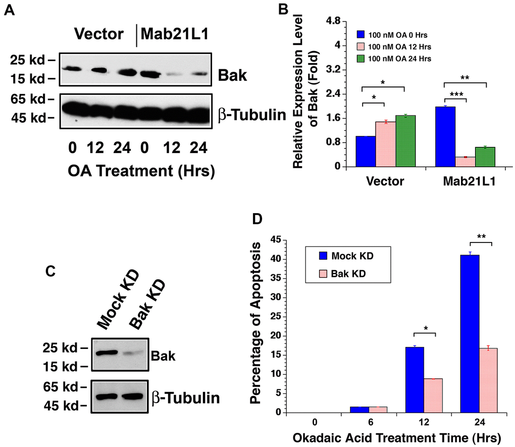
Figure 7.Dynamic change of Bak levels in vector-αTN4-1 and MAB21L1-αTN4-1 cells in the absence and presence of 100 OA treatment and effect of Bak level on OA-induced apoptosis of αTN4-1 cells. (A, B). Dynamic change of Bak levels in vector-αTN4-1 and MAB21L1-αTN4-1 cells in the absence and presence of 100 OA treatment. Both vector-αTN4-1 and MAB21L1-αTN4-1 cells were grown to about 90% confluence and then subjected to 100 nM OA treatment for 12 and 24 hrs. Thereafter, the cells were harvested for extraction of total proteins which were used for analysis of Bak expression by Western blot analysis (A). Quantitative results of the Bak protein expression levels were analyzed by Image J software (B). Note that in the MAB21L1-αTN4-1 cells, the Bak protein expression level was much higher than that in the vector-αTN4-1 cell clone in the absence of 100 nM OA treatment. During OA treatment for 12 and 24 hrs, however, Bak protein was upregulated in vector-αTN4-1 cells. In contrast, in MAB21L1-αTN4-1 cells, Bak protein was significantly degraded. N=3. *p<0.05, ** p<0.01, *** p<0.001. (C, D) Effect of Bak level on OA-induced apoptosis of αTN4-1 cells. Vector-αTN4-1 cells were used as Bak knockdown with CRSPR/Cas9 technology (see Materials and Methods). Both mock and Bak KD clones were verified with Western blot analysis (C). The two types of cells were then subjected to 100 nM OA treatment for 12 and 24 hrs, and the apoptosis rate was determined with live/dead assays (D). N=3. *p<0.05, ** p<0.01.