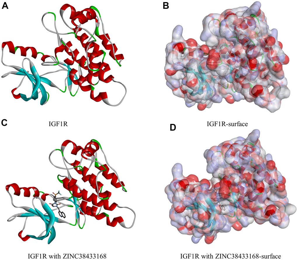
Figure 1.Molecular structure of IGF-1R. (A) Initial molecular structure. (B) Surface of binding area added. Blue represents positive charge, and red represents negative charge. (C) Molecular structure after IGF-1R and ZINC38433168 binding. (D) Surface of IGF-1R and ZINC38433168 binding area added. Blue represents positive charge, and red represents negative charge.